
Skin Cancer Photos – Common Types and Tips for Early Detection

In this article
Skin cancer is an increasingly common health problem today. In particular, sun exposure and harmful ultraviolet (UV) rays can significantly increase skin cancer risk. Raising public awareness and emphasizing early detection are therefore very important. This article, entitled "Skin Cancer Photos," provides a visual guide to recognizing and understanding the different types and signs of skin cancer.
In this article, we will examine photographs of skin cancers, particularly melanoma , as well as basal cell carcinoma and squamous cell carcinoma. We will also provide information about skin cancer signs, the importance of early detection, and protective measures.
What Are the Most Common and Rarest Skin Cancers?
Skin cancers differ in frequency and prevalence. Here are the most common and rarest types:
The most common skin cancer types:
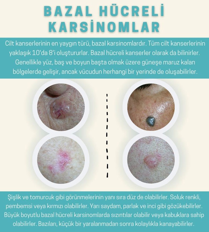
- Basal cell carcinoma (BCC): Accounting for approximately 70-80% of skin cancers, this type arises from cells in the uppermost layer of skin. BCC generally grows slowly and rarely metastasizes—that is, it does not spread to other organs.
- Squamous cell carcinoma (SCC): Accounting for approximately 15-20% of skin cancers, this type arises from flat cells in the middle layer of skin. SCC grows faster than BCC and has a higher risk of metastasis.
The rarest skin cancer types:
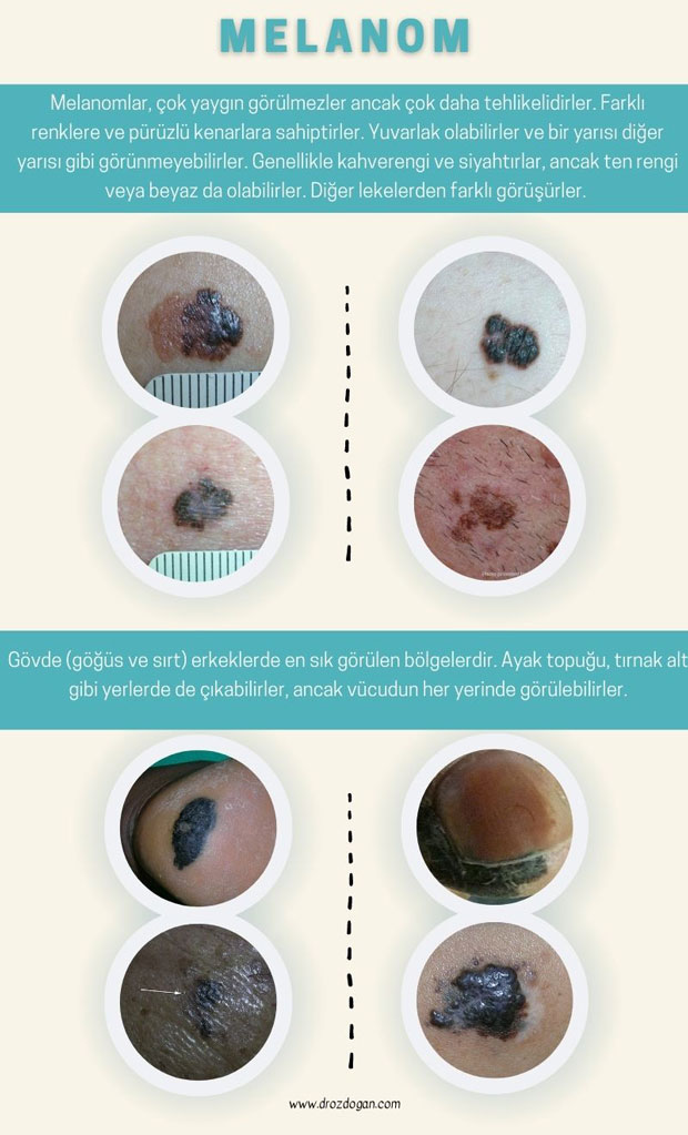
- Melanoma: Accounting for approximately 1-2% of skin cancers, this type arises from pigment-producing cells called melanocytes. Melanoma is more aggressive than other skin cancer types and more dangerous because of its high potential to metastasize.
- Merkel cell carcinoma: This rare skin cancer arises from Merkel cells in the skin's outer layer. It grows rapidly, has a high potential to metastasize, and requires early diagnosis and treatment.
- Kaposi sarcoma: This type arises from blood vessel and lymph node cells and is generally seen in people with weakened immune systems. Kaposi sarcoma is more common in people with HIV/AIDS and in immunosuppressed patients following organ transplantation.
Although some skin cancer types are rare, knowing the signs and risk factors is important. Early diagnosis and treatment improve the chances of successful outcomes.
Basal Cell Carcinoma Photos

*
Squamous Cell Carcinoma Photos

*
Malignant Melanoma Photos
*
Cutaneous Lymphoma and Kaposi Sarcoma Photos

*
Merkel Cell Carcinoma Photo

*
What Is Dermoscopy?
Dermoscopy (also called dermatoscopy ) is a diagnostic tool used to assess skin lesions and signs of skin cancer. A dermoscope provides magnified images of features such as the color, structure, and pattern of skin lesions and moles. This can help doctors assess the likelihood of skin cancer and detect it at an early stage.
Dermoscopes are divided into two main types: contact and noncontact. Contact dermoscopes touch the skin surface during use, whereas noncontact dermoscopes work without touching the skin and incorporate digital cameras and imaging technology.
The advantage of a dermoscope is that it allows better assessment of lesions and moles, providing an initial evaluation before more invasive procedures such as biopsy. However, consulting an experienced specialist is important, and suspicious lesions may require a more comprehensive evaluation and additional tests for a definitive diagnosis.
What Is the Difference Between Skin Cancer and a Mole?
Doctors frequently receive questions about skin cancers and moles, both in their professional and everyday lives. In this article, alongside important practical information, I want to draw particular attention to the sun's harmful effects and how moles should be monitored.
Skin cancer arises when cells in different layers of the skin grow uncontrollably.
A mole, also known as a nevus, is a common skin growth made up of pigment-producing melanocytes. Moles are a normal part of the skin, are generally harmless, and do not require treatment. Although moles can sometimes be cancerous, this is very rare. Cancerous moles are called melanomas and arise from pigment-producing melanocytes in moles. They can cause serious problems if not detected early.
The main difference between skin cancer and moles is that cancer develops through uncontrolled cell growth. Moles generally have a stable size and shape and usually do not change. Skin cancer often results from sun exposure, whereas moles may be related to genetic factors. Any change in a mole's size or shape should prompt a consultation with a specialist without delay.
Check Your Skin
Regular skin checks are important in determining whether a mole is cancerous
- Stand in front of a full-length mirror and inspect the skin for new or changing moles, spots, or lesions, checking the entire body, including the scalp, back, and soles.
- Use a hand mirror to examine areas that are difficult to see, such as the backs of the thighs or the back.
- Pay attention to each mole or spot's size, shape, color, and texture, and note any changes since the last check.
The Ugly Duckling Test
Another useful self-check technique is the ugly duckling test. This involves identifying any mole or spot that looks different from the others on the body.
Anyone who notices an unusual or changing mole or spot, or has concerns about a skin spot, should consult a specialist for professional assessment. Self-examination is an important step toward early detection of skin cancer.
When to See a Doctor
Consult a specialist if you develop a new mole or notice changes in an existing mole. Other changes to watch for include a mole or spot that bleeds or becomes itchy and painful.
Regular specialist skin checks are also important for people with a family history of melanoma, fair skin, a history such as sunburn, or a high risk of developing the disease.
In Summary: A Mole or Cancer?
Learning about skin cancer and moles and performing self-checks are important. A dermatological examination is needed to determine whether a mole is melanoma. Regular monitoring of dysplastic nevi and noticing skin changes are important for early skin cancer diagnosis. Self-examination, regular dermatological examinations, and sun protection measures can help prevent skin cancer.
Was this article helpful?
Your feedback helps us improve our content.




Reader comments
Share your comment
Your name and comment are published only after administrator approval. Your email address is never shown to visitors.
Be the first to comment.