
From a Ram’s Testicle to Modern Radiotherapy: How Invisible Rays Became a Safe Weapon

In this article
Radiotherapy’s First Brave and Dangerous Years, the Discovery of Fractionation, and the Invisible Ray’s Transformation into a Weapon
Wilhelm Röntgen discovered X-rays (day 81, 1895); Marie and Pierre Curie discovered radium (day 82, 1898). But these discoveries were not yet a treatment — merely two new phenomena emitting invisible rays. Turning them into a controlled, safe, effective cancer treatment required a separate, painful story. That story began astonishingly quickly: just weeks afterRöntgen announced his discovery, in early 1896, physicians around the world began applying X-rays to tumors — often without knowing the dose, without protection, without any scientific basis, relying purely on trial-and-error courage. Emil Grubbe in Chicago, Victor Despeignes in Lyon, Leopold Freund in Vienna: some saw dramatic improvements in skin cancers; others caused terrible radiation burns, necrosis, and secondary cancers in their patients and their own hands. The early years were an uncontrolled period generating both hope and tragedy. The problem was that a large radiation dose given at once killed the tumor while also burning and destroying healthy tissue. The solution came from an unexpected place: a ram’s testicle. At the Institut Curie in Paris, radiobiologist Claudius Regaudnoticed something extraordinary in experiments on ram testes: a single large radiation dose could not halt sperm production, while severely burning the scrotal skin, but giving the same total dose divided over several dayspermanently stopped sperm production while sparing the skin. This marked the birth of modern radiotherapy’s most fundamental principle: fractionation , dividing the dose into small parts spread over time. Regaud’s brilliant student Henri Coutardapplied the principle to head and neck cancers and, in Paris in 1922, showed the world patients with advanced laryngeal cancer cured by radiation. Today, on day 83 of the series, we examine radiotherapy’s transformation from blind courage into a precise science.
The first tumor treatments, months after Röntgen’s discovery
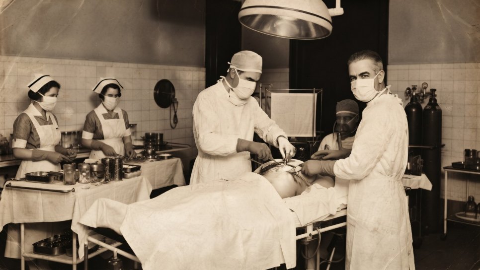
The medical imaging value of X-rays was clear (day 81); more surprising was how quickly their use for treatment began. Röntgen announced his discovery in December 1895, and in the first months of 1896 physicians in different parts of the world were already applying X-rays to skin diseases and tumors. An ironic historical fact: X-rays were initially used more for treatment than diagnosis — contrary to a modern reader’s intuition.
⚛ RADIOTHERAPY’S FIRST BOLD EXPERIMENTS (1896)
- Emil Grubbe (Chicago, 29 January 1896): A vacuum-tube manufacturer and medical student, Grubbe treated a breast cancer patient with X-rays on Dr. Ludlam’s suggestion — the first known therapeutic X-ray application in America. Grubbe’s own hands sustained severe radiation damage over the years; he underwent numerous amputations.
- Victor Despeignes (Lyon, July 1896): The French physician treated a stomach cancer patient with X-rays. Interestingly, based on the hypothesis that “the dose should not be too high, and healthy tissue must be protected”, Despeignes tried a bi-fractionated approach, with two divided doses — a very early, intuitive precursor of fractionation.
- Leopold Freund (Vienna, 1896-97): The Viennese dermatologist treated a hairy nevus, a birthmark, with X-rays. The weak X-ray tubes of the time delivered very low doses, so Freund had to treat daily, at a low dose, for weeks until obtaining an effect. The first radiotherapy treatments were therefore already fractionated out of technical necessity. Freund is considered a founder of modern medical radiology and radiotherapy.
- Danlos (Hôpital Saint-Louis, Paris, 1901): The first therapeutic use of radium — “brachytherapy” for skin, applying the radioactive source directly to the skin.
A technical limitation: The biggest limitation of the early period was the low energy produced by X-ray tubes, the Crookes tubes — only a few tens of kilovolts. These “soft” X-rays could not penetrate deeply into the body, so early treatments were mostly confined to lesions on the skin surface: skin cancers, lupus, eczema, and nevi. Reaching deep tumors only began to become possible in 1913 with William Coolidge’s more powerful, controllable “Coolidge tube.” The limits of technology also defined the limits of clinical imagination.
The central dilemma of early radiotherapy
In the first decade of the 1900s, radiotherapy faced a central problem. Physicians tended to deliver the highest possible radiation dose in one sitting to destroy the tumor. The logic was simple: “the more radiation, the more dead tumor”. But the result was often disastrous. Even if a single large dose killed the tumor, it severely burned healthy tissue along its path, particularly the skin, caused necrosis or tissue death, and left nonhealing wounds and permanent damage. The tumor might shrink, but the treatment itself seriously harmed the patient.
The problem was biological, and no one yet understood its mechanism. Its solution would also come from a biological observation, arising from a wholly unexpected experimental subject.
Claudius Regaud and the foundational experiment of radiobiology
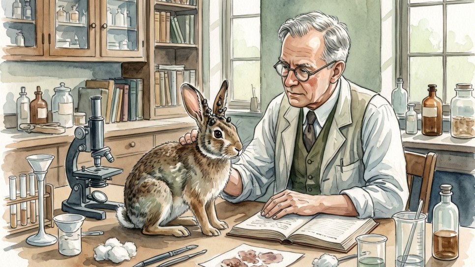
Claudius Regaud’s ram-testis experiments · Birthplace of a fundamental principle of modern radiotherapy
Claudius Regaud (1870-1940) was the radiobiologist heading the biology and medicine division of Marie Curie’s Radium Institute. To understand how cells respond to radiation, he sought tissue containing continuously dividing cells and chose the ram’s testicle, because sperm-producing cells, spermatogonia, are among the body’s fastest-dividing and therefore most radiation-sensitive cells.
Regaud found something striking: a single large radiation dosecould not permanently stop the ram’s sperm production, yet delivering that dose caused unbearable burns to the scrotal skin. In contrast, dividing the same total dose over several days into successive small doses permanently stopped sperm production while sparing the scrotal skin.
The result was revolutionary: a divided, fractionated dose was both more effective and safer than a single large dose. Healthy tissue could repair itself between doses, while rapidly dividing target cells, such as tumor cells, could not perform this repair and were cumulatively destroyed.
📐 THE FRACTIONATION PRINCIPLE — RADIOTHERAPY’S CORNERSTONE
The principle discovered by Regaud remains the foundation of all modern radiotherapy: dividing the total radiation dose into small “fractions” spread over days. The rationale rests on the difference in repair between healthy and tumor tissue.
Healthy cellscan repair their DNA damage and survive during the interval between two fractions, usually 24 hours. Tumor cells , by contrast, are more sensitive to radiation because they divide faster, and their DNA repair mechanisms are often impaired, preventing recovery between fractions. They suffer cumulatively greater damage with each fraction. Small daily doses spread over weeks can thus gradually destroy the tumor while protecting healthy tissue — a “therapeutic window” opens.
SINGLE DOSE vs FRACTIONATED DOSE — SAME TOTAL, DIFFERENT OUTCOME
⚠ SINGLE LARGE DOSE — early period
Kills the tumor, but healthy tissue burns without a chance to repair. Necrosis, nonhealing wounds, permanent damage. The patient is harmed by treatment.
✓ FRACTIONATED DOSE — Regaud-Coutard
The same total dose in small daily fractions spread over weeks. Healthy tissue repairs each night; the tumor is cumulatively destroyed. Cure with acceptable damage.
From the basement X-ray unit to clinical radiotherapy
The person who applied Regaud’s laboratory discovery to actual patients was his brilliant colleague at Institut Curie, Henri Coutard (1876-1950). Coutard’s story is interesting: during the First World War he headed the radiology service of a military hospital on the Eastern Front, while also serving in Marie Curie’s mobile X-ray units, the “petites Curies” (day 82). After the war, he joined Institut Curie and began working in an X-ray unit in its basement.
Coutard applied Regaud’s fractionation principle to head and neck cancers, especially cancers of the larynx and pharynx. He spread treatment courses over weeks, delivered small daily doses, and meticulously observed skin and mucosal reactions. He systematically described radiation side effects, introducing the terms “radioepidermitis” for radiation-induced skin peeling and “radioepithelitis” for the radiation reaction of the oral and pharyngeal mucosa. He also made a crucial observation: different tumors have different sensitivities to radiation — they do not respond identically to the same dose. This formed the basis of the modern concept of “radiosensitivity.”
Six laryngeal cancer patients · The birth of clinical radiotherapy
At the International Congress of Otology in Paris in 1922, Regaud, Coutard, and their colleague Hautant presented a striking finding to the world: six patients with advanced laryngeal cancerhad been treated with fractionated radiotherapy and become disease-free .
This was the first demonstration in history that radiation treatment could control an advanced cancer without surgery. Until then, laryngeal cancer had usually been fatal or required radical surgery with devastating effects on voice and swallowing. Coutard’s patients were cured while retaining their organs and functions.
For this reason, the beginning of clinical radiotherapy is generally dated to 1922.
Coutard continually refined his approach. Initially, he delivered large daily doses at a low dose rate to small tumors. When he reduced the daily dose for larger tumors, he found that a low dose rate was no longer needed. This led to the “simple fractionation” technique: small fraction sizes at a relatively high dose rate. Around 1930, leading radiotherapists in Zurich (Schinz), Vienna (Holzknecht), and other centers adopted it.
The First International Congress of Radiology
In London in 1925, at the First International Congress of Radiology, Claudius Regaud formally presented the fractionation principle and Institut Curie’s “French school” approach to the international scientific community. This began the acceptance of fractionated radiotherapy as a global standard. Paris’s Institut Curie, the institution born from Marie Curie’s discovery of radium, had also become the center of world radiotherapy. Its “French school,” formed by Regaud, Coutard, Antoine Lacassagne, and others, defined the basic clinical principles of modern radiation oncology in the 1920-1930s.
⚠ THE PIONEERS’ PRICE — TRAGEDIES OF EARLY RADIOTHERAPY
Radiotherapy’s birth years brought not only cures but also a heavy price. Because radiation hazards were unknown, early physicians and technicians worked without protection. Emil Grubbedeveloped severe radiation damage to his hands after working with X-rays for years and underwent repeated operations and amputations. Marie Curie herself (day 82) died of radiation-related aplastic anemia. Many early radiologists developed radiation dermatitis on their hands and later skin cancer.
Patients suffered tragedies too. In the early years, when doses were unknown, excessive radiation caused necrosis, chronic wounds, bone damage, and secondary cancers. These painful experiences, the price paid by pioneers and patients, led to the emergence of modern radiation safety science — radiobiology, dose limits, protective equipment, and the ALARA principle, “as low as reasonably achievable.” Fractionation itself was one of the greatest products of this search for safety.
More powerful rays, deeper tumors
The biggest technical limitation of early radiotherapy was its low beam energy: soft X-rays could not reach deep tumors. This limitation was overcome in the middle of the 20th century:
⚛ THE EVOLUTION OF RADIOTHERAPY TECHNOLOGY
The Coolidge tube (1913): William Coolidge’s controllable, higher-energy X-ray tube became the standard for “kilovoltage” treatment, but penetration remained limited.
Cobalt-60 units, the “cobalt bomb” (1950s): High-energy gamma rays from the artificial radioactive cobalt-60 isotope could reach deep tumors. Cobalt units spread radiotherapy worldwide. Relatively inexpensive and durable, they remained essential tools for decades, particularly in developing countries.
Linear accelerator (LINAC, from the 1950s onward): Devices that accelerate electrons to high speeds to generate high-energy X-rays or electron beams. They are today the standard for external-beam radiotherapy and have largely replaced cobalt units.
Modern techniques: Intensity-modulated radiotherapy (IMRT), image-guided radiotherapy (IGRT), stereotactic radiosurgery (SRS/SBRT), and proton therapy are all refined descendants of Regaud’s fractionation principle, applying it with millimetric precision while maximally protecting healthy tissue.
The institutionalization of radiotherapy in Turkey
Modern radiotherapy’s arrival in Turkey progressed alongside the Republic’s healthcare advances. During the first half of the 20th century, radium tubes and kilovoltage X-ray units were installed; from the middle of the 20th century, cobalt-60 devices were established in university hospitals and major urban hospitals. Linear accelerators subsequently spread. Today, IMRT, SBRT, and modern image-guided radiotherapy are routinely delivered at many centers across Turkey.
In contemporary oncology, radiation oncology is one of three main pillars alongside surgery and medical oncology. Organ-preserving chemoradiotherapy for head and neck cancer, directly descended from Coutard’s laryngeal patients in 1922; postoperative adjuvant radiotherapy for breast cancer; SBRT for prostate cancer: all are current extensions of the fractionation principle born from a ram’s testicle. A radiation oncologist at the table for every case discussed by multidisciplinary tumor boards is today’s expression of this 100-year lineage.
⚛ FROM BLIND COURAGE TO PRECISION SCIENCE
🔍 INFORMATION VERIFIED IN THIS ARTICLE
- Emil Grubbe: Chicago, 29 January 1896, first therapeutic X-ray use in a breast cancer patient in the USA, on Dr. Ludlam’s suggestion. Vacuum-tube manufacturer and medical student. Radiation damage to hands, amputations.
- Victor Despeignes: Lyon, 1896, stomach cancer; bi-fractionated approach.
- Leopold Freund (1868-1943): Vienna, 1896-97, hairy nevus; daily low-dose treatment for weeks. Considered a founder of modern medical radiology/radiotherapy.
- Danlos: Hôpital Saint-Louis, Paris, 1901, first therapeutic radium use, brachytherapy.
- Technical limitation: Early Crookes tubes: a few tens of kilovolts; “soft” X-rays could not penetrate deeply. Coolidge tube, 1913.
- Claudius Regaud (1870-1940): Radiobiologist at Institut Curie/Fondation Curie. Ram-testis experiments: a single large dose cannot stop sperm production and burns the scrotum; fractionated doses stop sperm production and spare the skin. The foundation of fractionation.
- Henri Coutard (1876-1950): Basement X-ray unit at Institut Curie. Radiology at a WWI military hospital on the Eastern Front plus Marie Curie’s petites Curies units. Fractionated head and neck radiotherapy. Terms “radioepidermitis” and “radioepithelitis.” Observation of differing radiosensitivity among tumors. Move to the Chicago Tumor Institute in 1930.
- Paris Congress of Otology, 1922: Regaud, Coutard, Hautant: 6 advanced laryngeal cancer patients disease-free after radiotherapy. Considered the beginning of clinical radiotherapy (1922).
- London, 1925: First International Congress of Radiology; Regaud presented fractionation.
- The French school: Institut Curie: Regaud, Coutard, Antoine Lacassagne; basic principles of modern radiation oncology in the 1920-30s.
- Technological evolution: Coolidge tube (1913) → cobalt-60 (1950s) → linear accelerator → IMRT/IGRT/SBRT/proton.
“Radium Girls”: Glowing Paints, an Industrial Scandal, and the Birth of Occupational Health
The discovery of radium (day 82) and radiation’s medical power (day 83) also triggered a public craze for radium in the early 20th century. The “miracle element” was added to cosmetics, drinking water, toothpaste, and even glow-in-the-dark watch dials. In factories in New Jersey and Illinois, young women painted watch dials by hand with radium paint and licked their brushes to sharpen the tips, swallowing more radium with every lick. The result was one of the most shocking industrial health tragedies in medical and legal history: the “Radium Girls,” whose jawbones disintegrated and who developed bone cancer. Their lawsuit helped bring about modern occupational health and safety laws, radiation protection standards, and the concept of corporate responsibility. On day 84, we will examine this tragedy behind the glow.
Author’s note: The Coutard quotation in this article is a conceptual summary of his work in the 1920s from the perspective of modern radiobiology. Priority in early radiotherapy is historically debated among many names, including Grubbe, Despeignes, Freund, Voigt, and Schiff. Most early attempts were one-off, nonsystematic interventions; what made radiotherapy a science was Regaud and Coutard’s systematic, fractionation-based approach. The biological basis of fractionation — healthy tissue repair, tumor radiosensitivity, and the “4R”: repair, redistribution, reoxygenation, and repopulation — can be addressed in detail in later installments on radiobiology. Day 84 will cover the “Radium Girls” and occupational health; later installments will examine radiobiology’s molecular foundations.
Sources and Further Reading
- Regaud C, Ferroux R. Discordance des effets de rayons X, d'une part dans la peau, d'autre part dans le testicule, par le fractionnement de la dose. Comptes Rendus de la Société de Biologie 1927;97:431-434. (Original publication of the fractionation principle.)
- Coutard H. Roentgen therapy of epitheliomas of the tonsillar region, hypopharynx and larynx from 1920 to 1926. American Journal of Roentgenology 1932;28:313-331. (Coutard’s clinical series.)
- Thames HD, Hendry JH. Fractionation in Radiotherapy. London: Taylor & Francis, 1987. (A classic examination of fractionation history and radiobiology.)
- Bernier J, Hall EJ, Giaccia A. Radiation oncology: a century of achievements. Nature Reviews Cancer 2004;4(9):737-747. (A century of radiation oncology history.)
- Connell PP, Hellman S. Advances in Radiotherapy and Implications for the Next Century: A Historical Perspective. Cancer Research 2009;69(2):383-392.
- Coutard H — A Pioneer of Fractionated Radiotherapy Regimens. PubMed Central Biographical review (PMC11471863). (Coutard’s life and contributions.)
- The ASCO Post. History of Radiation Oncology in the United States. 25 June 2022.
- History of Radiation Biology. Springer Nature, 2023. (The early period: Freund, Despeignes, Regaud, Coutard, Lacassagne.)
- Mould RF. A Century of X-rays and Radioactivity in Medicine. Bristol: Institute of Physics Publishing, 1993.
- Mukherjee S. The Emperor of All Maladies: A Biography of Cancer. New York: Scribner, 2010. (The birth of radiation oncology; Turkish edition: İmparatorluğun Kralı, Domingo Publishing.)
- Turkish Society for Radiation Oncology (TROD). A review of the history of radiation oncology in Turkey.
This article is day 83 of the Cancer History in 365 Days Series. The birth of radiotherapy — turning invisible rays into a controlled cancer treatment after Röntgen’s discovery of X-rays (day 81) and the Curies’ discovery of radium (day 82) — is the foundation of radiation oncology, one of modern oncology’s three main pillars. Claudius Regaud’s fractionation principle and Henri Coutard’s clinical application transformed radiotherapy from dangerous trial and error into a precise science that destroys tumors while sparing healthy tissue. This principle remains the basis of all modern radiotherapy, including IMRT, SBRT, and proton therapy. Early radiotherapy’s tragic price — radiation damage to pioneering physicians and overdose complications in patients — led to modern radiation safety science. This sensitive subject is presented in balanced, informative language for readers anxious about radiation. The article takes a historical, popular perspective and provides no treatment guidance. All dates, names, and scientific information were verified using Regaud and Coutard’s original publications, the Bernier-Hall-Giaccia history in Nature Reviews Cancer (2004), the Connell-Hellman perspective in Cancer Research (2009), Springer’s history of radiation biology (2023), and modern academic sources.
Was this article helpful?
Your feedback helps us improve our content.


Reader comments
Share your comment
Your name and comment are published only after administrator approval. Your email address is never shown to visitors.
Be the first to comment.