
How Did Marcello Malpighi Change the History of Cancer with the Microscope?

In this article
Day 58 (The History of Cancer in 365 Days Series)
The first discovery of the invisible: the medical revolution Malpighi, the father of modern histology, embryology and pathology, built behind a lens.
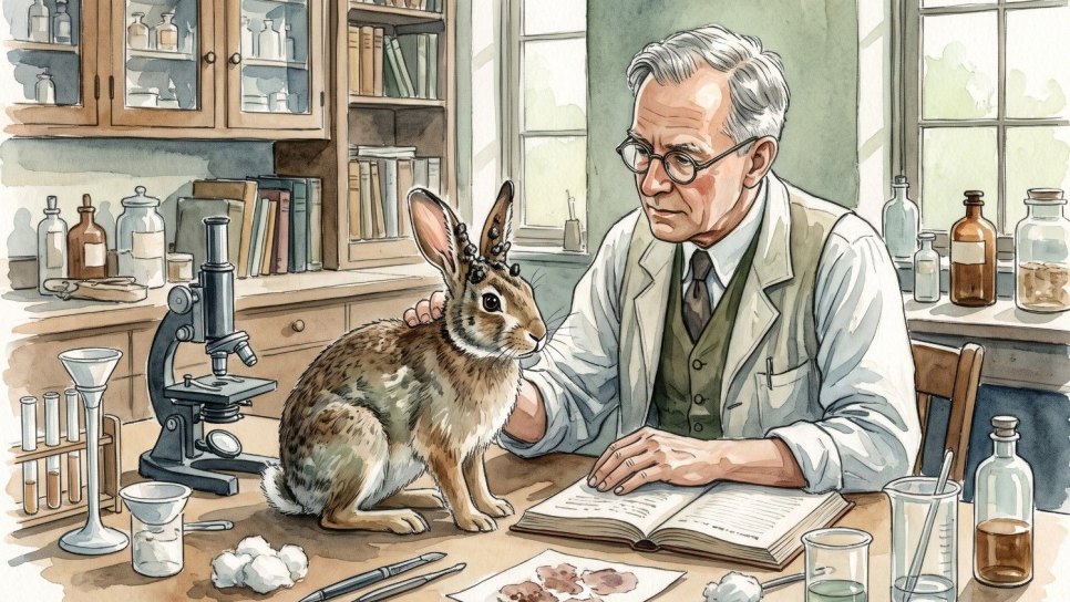
For centuries, the history of cancer had been confined by the naked eye. Physicians saw tumours and cut them out, yet could never understand why they spread or what lay deep within them. By turning the earliest microscopes towards human tissue, Marcello Malpighi proved that organs were not random masses of flesh but flawless works of architecture. The foundations of the disciplines of “biopsy” and “cellular analysis” used in pathology today were laid by the light that passed through Malpighi’s first lens. Malpighi did more than see tissues: by discovering capillaries, he completed the missing link in the theory of blood circulation and carried medical history beyond the age of “observation” into the age of “analysis”.
Born in Crevalcore, Italy, in 1628, Marcello Malpighiis regarded as one of the greatest rebels in medical history. Having studied medicine and philosophy at the University of Bologna, Malpighi rejected medical education based on memorising ancient books and sought truth in the laboratory and in experiments on living animals.
1. From a mass of flesh to flawless architecture: the birth of histology
While serving as a professor at the universities of Bologna, Pisa and Messina, Malpighi targeted medicine’s greatest blind spot: the internal structure of organs. Until then, Galen’s and Hippocrates’ “humoral theory” had regarded organs as spongy masses of tissue. When Malpighi directed his lenses at these masses, he recognised a startling truth.
He showed that every organ contained microscopic chambers, glands and ducts. He was the first to describe the tactile endings in the skin and the kidney’s “Malpighian Corpuscles”.
By discovering the capillary vessels, he scientifically filled the gap of the “transition between arteries and veins” in William Harvey’s theory of blood circulation.

By observing pulmonary alveoli for the first time, Malpighi established that respiration was an interaction between air and blood rather than an “exchange of fluids”. These studies gave birth to the science that underpins modern physiology and pathology: Histology .
2. The dawn of embryology: life in the egg
Malpighi placed not only adult tissues but also the beginning of life under the microscope. He became one of the first scientists to examine chicken embryos at very early stages. In his work published in 1673, De Formatione de Pulli in Ovo (On the Formation of the Chick in the Egg), he documented in detail how a living organism is built.

Above, "Formazione pulli...", 1687. De formasyon pulli in ovo. Chick embryology.
Preformation Theory and the Modern Legacy
By observing embryonic development through the microscope, Malpighi identified the changes that organs undergo during development. His drawings and observations were used for centuries as fundamental references in understanding embryology and developmental biology. He was the first genius to prove that life takes shape not “spontaneously” but according to a microscopic plan.
3. Mapping cancer’s pathways: the secret of the lymph nodes
Malpighi’s greatest contribution to the history of cancer was his work published in 1689, De structura glandularum conglobatarum . In the terminology of the time, “conglobate glands” were Lymph Nodes.
Wilhelm Fabry (Day 49) had sensed the metastatic power of the axillary glands in breast cancer, but Malpighi illuminated these glands’ internal structure . He showed that they were not simple swellings but complex filters containing a network. The anatomical blindness that had prevented us from understanding why cancer gets caught at these “stations” as it spreads became a thing of the past through Malpighi’s lens.
International recognition and an ethical legacy
Malpighi’s work was so groundbreaking that England’s Royal Society of London accepted him as an honorary member and began publishing his works. Malpighi, who also served as chief physician to Pope Innocent XII, was known as an ethical and gentlemanly scientist who, despite his scientific success, bore no grudge against colleagues who opposed him.
Seeing is the first step in restoration
Marcello Malpighi bequeathed to us the most fundamental ability needed to provide “precise guidance” in oncology: the ability to “see”. By looking inside tissues, he discovered not only organs but also the microscopic pathways of healing. This journey, extending from capillaries to lymphatic filters, is the oldest philosophical counterpart of the discipline we now call “molecular targeting”.
The philosophy of biological restoration begins with knowing the normal structure of a tissue and understanding where that structure has broken down (pathology). The curious and gentlemanly stance Malpighi displayed in the laboratories of the 17th century continues to illuminate today’s digital pathology and artificial intelligence analyses. Healing means recognising the perfect balance in nature’s smallest detail and being able to rebuild that balance through science.
Historical and Academic Sources
- 1. Adelmann HB. Marcello Malpighi and the Evolution of Embryology. Cornell University Press, 1966. (A 5-volume reference work).
- 2. Motta PM. "Marcello Malpighi and the foundations of functional microanatomy." The Anatomical Record, 1998;253(1):10-15.
- 3. Malpighi M. De viscerum structura exercitatio anatomica. (1666) and De Formatione de Pulli in Ovo. (1673).
- 4. Çelik M. "Lenfatik Sistemin Tarihçesi ve Malpighi'nin Katkıları." Lokman Hekim Dergisi, 2023.
Was this article helpful?
Your feedback helps us improve our content.


Reader comments
Share your comment
Your name and comment are published only after administrator approval. Your email address is never shown to visitors.
Be the first to comment.