Liver Hemangioma or Metastasis? Telling Them Apart: Why Dynamic Liver MRI Is Ordered, What It Shows
In this article
Liver Hemangioma or Metastasis? Telling Them Apart: Why Dynamic Liver MRI Is Ordered, What It Shows
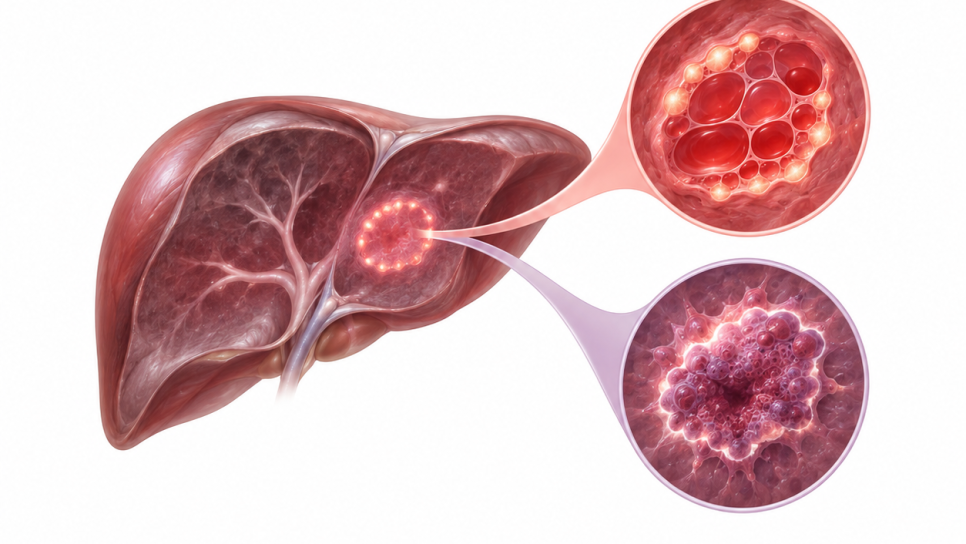
A liver lesion found by chance on ultrasound or CT, especially in someone with a history of cancer, raises the question "hemangioma or metastasis?" A hemangioma is a very common, benign vascular lesion; a metastasis is cancer from another organ that has settled in the liver. Follow-up and treatment are completely different. Dynamic liver MRI can often settle the question by looking at the lesion's T2 brightness, diffusion and how injected contrast spreads through it over time; but it is not conclusive for every lesion. This article explains what MRI looks at, what each finding means and where the pitfalls are.
- The question: Is an uncertain liver lesion a benign hemangioma or a metastasis? The answer changes the treatment plan; a wrong read can mean unnecessary cancer treatment or a delayed diagnosis.
- Why dynamic MRI? On top of T2 and diffusion images, it follows how the lesion enhances after contrast in the arterial, portal venous and delayed phases. A typical hemangioma fills in from the edge toward the center; a metastasis usually enhances as a ring and lets go of contrast in the delayed phase.
- What it cannot do alone: No single sign decides. T2, diffusion and the dynamic phases are read together; it gets harder with small lesions (under about 2 cm), atypical lesions, after chemotherapy and with hypervascular primaries (neuroendocrine tumor, kidney, thyroid, melanoma).
- What to do: With a cancer history, the MRI should be interpreted together with your oncologist; if doubt remains, contrast-enhanced ultrasound, short-interval follow-up or biopsy is discussed at a tumor board.
What are a hemangioma and a metastasis?
A hemangioma (cavernous hemangioma) is one of the most common benign tumors of the liver; it is made of blood-filled vascular spaces and causes no symptoms in most people. It is usually found by chance on imaging done for another reason. By size it is classed as small (about 1-2 cm), typical (2-10 cm) or giant (over 10 cm).
A liver metastasis is the spread to the liver of cancers from other organs, such as colon and rectum, breast, lung, pancreas, neuroendocrine tumors, kidney, thyroid and melanoma. Liver metastases are many times more common than primary liver tumors; they are often multiple, but can be single.
In short: a hemangioma is the liver's own benign vascular lesion; a metastasis is a cancer from elsewhere that has settled in the liver. One does not turn into the other; the problem is that they can look alike on images.
What is dynamic liver MRI?
Dynamic (contrast-enhanced) liver MRI includes, in one exam, sequences without contrast followed by images taken in rapid succession after a gadolinium-based contrast agent is injected into a vein. "Dynamic" refers to watching how the contrast spreads through the liver over time. The radiologist looks not at one image but at the pattern these images form together.
| Image / phase | What is seen? | Why it matters |
|---|---|---|
| T2-weighted | Fluid content of the lesion; a hemangioma is very bright, a metastasis usually mildly to moderately bright | First distinguishing clue; but cystic and neuroendocrine metastases can also be bright |
| Diffusion (DWI/ADC) | Restriction of water movement; a metastasis is expected to show restriction and low ADC | Helps even when contrast cannot be given |
| Arterial phase | First arrival of contrast through the vessels (first seconds) | Peripheral nodules in a hemangioma; a thin ring or intense enhancement in a metastasis |
| Portal venous phase | About the first minute | In a hemangioma filling advances toward the center; in a metastasis the center does not take up contrast |
| Delayed phase (a few minutes later) | Whether the lesion keeps the contrast | A hemangioma retains contrast; a metastasis usually releases it (washout) |
| Hepatobiliary phase (special agent only, about 20 min) | Whether liver cells take up the agent | Strong for showing small metastases; not enough alone to separate hemangioma from metastasis |
Figure 1 · Typical enhancement pattern on dynamic MRI
Simplified look of a hemangioma and a metastasis across four images
Which finding points to what?
The table below summarizes the typical appearances. No single row makes the diagnosis; the radiologist reads them all together and relates them to the patient's cancer history.
| Finding | Hemangioma | Metastasis | Note |
|---|---|---|---|
| T2 signal | Very bright ("light-bulb" sign); close to fluid | Usually mildly to moderately bright; mild brightness in 95% of colorectal metastases | Neuroendocrine and cystic metastases can be bright on T2 |
| Diffusion / ADC | High ADC; restriction usually absent or weak | Restriction, low ADC (87% in colorectal) | ADC values vary with scanner and b value |
| Arterial phase | Peripheral, discontinuous, nodular enhancement; as bright as the blood pool (aorta) | Hypovascular: thin ring or no enhancement; intense enhancement with hypervascular primaries | A small flash-filling hemangioma can mimic a hypervascular metastasis |
| Portal venous / delayed phase | Progressive fill-in toward the center; full or partial filling in the delayed phase, no washout | Ring stays prominent, center dark; washout in the delayed phase | In a giant hemangioma the center may never fill (fibrosis, thrombosis) |
| Hepatobiliary phase (gadoxetate) | Usually dark; "extended washout" may be seen on late images | Dark "hole"; a "target" look in 47% of colorectal lesions (70/150) | Not reliable alone for telling them apart |
| Number and context | Often single or a few; no symptoms | Often multiple; a known cancer history is critical | A single lesion does not exclude metastasis |
The table is compiled from summaries of Radiopaedia, StatPearls, van der Reijd et al. (2025) and related studies. The colorectal figures come only from colorectal metastasis series; other primaries may differ.
Figure 2 · Mean ADC values (single center)
Mean ADC by lesion type (×10⁻³ mm²/s); 134 lesions, 1.5 T, b = 50/400/800. Bars show this series only.
These values come from one center's series with one scanner and b value set (48 hemangiomas, 31 metastases). In another series of colorectal metastases the mean ADC was 1.31; values differ from center to center and there is no universal cut-off.
Why is dynamic MRI ordered?
When a liver lesion stays uncertain on ultrasound or CT, MRI is often one of the best tests to answer the question. In the American College of Radiology (ACR) 2020 criteria, for an indeterminate lesion over 1 cm in a normal liver in a patient with no suspected cancer, MRI without and with contrast is rated "usually appropriate"; multiphase CT is rated the same. The reasons can be listed as follows:
- Characterization: Looking at the same lesion over time is one of the strongest ways to separate a hemangioma from a metastasis that look alike on a single slice.
- Cancer history: In a patient with known cancer, every new liver lesion raises the chance of metastasis; the cost of a wrong call (unneeded chemotherapy or delayed treatment) is high.
- Small lesions: In lesions under 1-2 cm the patterns do not become clear; a flash-filling hemangioma can mimic a hypervascular metastasis.
- Other liver disease: Against a fatty liver, small colorectal metastases have been shown to be detected more sensitively on hepatobiliary-agent MRI than on CT; in cirrhosis, a small flash-filling hemangioma can mimic HCC.
- Staging and planning: If a metastasis is confirmed, showing its number and location affects the decision on surgery or ablation; MRI is considered more sensitive than CT for detecting metastases.
Figure 3 · Typical features of colorectal metastasis: before and after chemotherapy
Share of lesions showing the feature on gadoxetic acid MRI (%); 258 patients, 722 unique lesions. Bars show only these four criteria.
The six typical features: hypovascular, dark on unenhanced T1, arterial rim enhancement, no uptake in the hepatobiliary phase, mild T2 brightness and diffusion restriction. Because the typical look fades after chemotherapy, telling lesions apart can get harder in treated patients. The series is retrospective and single-center.
Pitfalls and limits
- Flash-filling hemangioma: Up to 16% of hemangiomas enhance quickly and uniformly in the arterial phase; most are under 2 cm and can mimic a hypervascular metastasis (neuroendocrine tumor, kidney, thyroid, melanoma). The clue is that contrast stays in line with the blood pool and there is no washout.
- Misleading "washout" with a hepatobiliary agent: A hemangioma can also look dark on late images with this agent; one study described this as an "extended washout sign". That is why the hepatobiliary phase is not used alone to tell them apart.
- Change after chemotherapy: In colorectal metastasis, mild T2 brightness fell from 95% to 79% and diffusion restriction from 87% to 65%; in a treated patient a metastasis may not look typical.
- Atypical hemangioma: Giant or degenerated (fibrosis, thrombosis, calcification) hemangiomas may not show the typical fill-in pattern and can be confused with a malignant lesion.
- Metastases bright on T2: Cystic and neuroendocrine metastases can be bright on T2 and may show diffusion restriction; looking at T2 alone misleads.
- If doubt remains: Contrast-enhanced ultrasound, Tc-99m-labeled red blood cell scintigraphy (SPECT), a short-interval follow-up MRI or biopsy are discussed at a tumor board.
Is the contrast agent safe?
- MRI contrast contains gadolinium; it differs from the iodinated contrast used in CT.
- The main concern is the rare nephrogenic systemic fibrosis (NSF) in severe kidney failure (eGFR below 30, dialysis); the risk has been linked especially to older agents. With macrocyclic (ACR group II) agents the risk is considered extremely low.
- That is why centers often ask about kidney function (creatinine/eGFR); the radiology team decides the suitable agent and dose. If contrast cannot be used, non-contrast MRI with diffusion can be complementary.
- Tell the team before the exam if you are pregnant or breastfeeding, or have a drug allergy or kidney disease.
- Ask when booking: the request "dynamic liver MRI" or "contrast-enhanced upper abdomen MRI"; if the hepatobiliary (late) phase is requested, the exam runs about 20 minutes longer. Which contrast agent is used varies by center.
- Bring previous images: ultrasounds, CTs and old MRIs (CD and report); how the lesion has changed over time is very valuable for the differential diagnosis.
- Kidney and allergy information: Have your latest creatinine/eGFR result and any drug allergies with you; whether it is required depends on the center's protocol.
- The decision is not made on one report: With a cancer history, the radiology report is reviewed together with your oncologist; when in doubt, a tumor board is recommended. If a metastasis is confirmed, molecular tests may be needed on the biopsy sample (what is next-generation sequencing (NGS)?, in Turkish).
- Reimbursement: Reimbursement conditions for contrast MRI were not verified in this article; ask your center.
Questions you can ask your doctor
- Which features of my lesion point toward hemangioma and which toward metastasis?
- Given my cancer history, is a contrast-enhanced (dynamic) MRI needed, and is a hepatobiliary agent preferred?
- Do my kidney function and allergies pose a problem for contrast?
- If the result is not certain, what is the next step: short-interval follow-up, contrast-enhanced ultrasound or biopsy?
- Does the report clearly state the number and location of all my lesions?
Common misconceptions
- "A liver lesion means cancer." No. Hemangiomas and cysts are very common; in someone with no cancer history an uncertain lesion is often benign, but with a cancer history the odds change and MRI clarifies it.
- "If MRI shows washout, it is a metastasis." No. Reading washout depends on the phase, the agent and the lesion type; with a hepatobiliary agent a hemangioma can also darken on late images.
- "Dynamic MRI always gives a definite diagnosis." No. In small, atypical and post-chemotherapy lesions, uncertainty can remain; then another method or follow-up is needed.
Frequently Asked Questions
- What is the difference between a hemangioma and a metastasis in the liver?
- A hemangioma is a benign vascular lesion of the liver; a metastasis is cancer from another organ that has settled in the liver. On MRI, a hemangioma is very bright on T2 and fills with contrast from the edge toward the center; a metastasis usually enhances as a ring and releases contrast in the delayed phase.
- Why is dynamic liver MRI ordered?
- To establish what an uncertain lesion is. How contrast spreads over time, together with T2 and diffusion findings, helps separate a hemangioma from a metastasis. It is especially valuable with a cancer history, a small or atypical lesion and other liver disease.
- Can the two be told apart without contrast?
- A typical hemangioma can often be recognized with T2 and diffusion; when contrast cannot be given, MRI with diffusion helps. But for small, atypical lesions and with a cancer history, a contrast-enhanced exam can be decisive.
- What happens if we are still unsure after MRI?
- Options such as contrast-enhanced ultrasound, red blood cell scintigraphy, a short-interval follow-up MRI or biopsy are weighed for each patient at a tumor board.
Glossary
- Hemangioma
- A benign liver lesion made of blood-filled vascular spaces.
- Metastasis
- Cancer from another organ that has settled in the liver.
- Dynamic MRI
- MRI taken in successive phases before and after contrast is given.
- T2-weighted image
- An MRI sequence that shows fluid content as bright; very bright in a hemangioma ("light-bulb" sign).
- Diffusion (DWI) / ADC
- Shows restriction of water movement in tissue; a low ADC means restriction.
- Arterial / portal / delayed phase
- The time windows in which contrast arrives from the vessels into the liver in turn.
- Washout
- Faster clearing of contrast from a lesion than from the surrounding tissue.
- Hypervascular
- A vessel-rich lesion that enhances more than the surrounding liver in the arterial phase.
- Flash-filling hemangioma
- A hemangioma that enhances quickly and uniformly in the arterial phase, usually under 2 cm.
- Hepatobiliary phase
- The phase taken about 20 minutes after a special agent that liver cells take up; it sharpens the difference between metastasis and normal tissue.
DROZDOGAN Academy Commentary
In separating hemangioma from metastasis, dynamic MRI is less about reading one sign than reading a pattern; it is strongest with typical lesions and weakest with small, atypical and treated ones.
⚡ Why it matters
Lesions found by chance in the liver are very common; in a patient with cancer, each one can change the treatment plan. Telling them apart correctly and without a procedure reduces unnecessary biopsies and treatment.
✅ Strengths
T2, diffusion and dynamic phases complement each other; the typical fill-in pattern is distinctive for hemangioma; MRI is more sensitive than CT for finding metastases; a strong place in the ACR criteria for an uncertain lesion.
⚠️ Limits
Most figures come from retrospective, single-center series; ADC values vary with the scanner; the percentages are specific to colorectal metastasis; flash-filling hemangioma and post-chemotherapy metastasis make separation harder; the hepatobiliary phase cannot separate them alone.
🩺 In practice
In a patient with a cancer history, dynamic MRI is a reasonable first step for an uncertain lesion; if the result is still unclear, follow-up or biopsy is discussed at a tumor board. Have the MRI read together with previous images.
❓ Open questions
How much do artificial intelligence and radiomics tools improve the separation in small lesions? Are abbreviated MRI protocols enough for this question? What should standard criteria look like for post-chemotherapy lesions?
Conclusion: Dynamic liver MRI is a widely used method and, in most lesions, strong enough to separate a hemangioma from a metastasis. But it rests on the whole pattern, not one sign; in small, atypical and post-chemotherapy lesions uncertainty can remain, and the decision should be made at a tumor board.
- This article was prepared in November 2026.
- The article uses a summary of some variants of the ACR Appropriateness Criteria (2020), the Radiopaedia and StatPearls reviews and the abstracts of the studies listed; the full texts of all studies were not examined directly. The ACR variants for patients with a cancer history were not verified and are not described.
- The figures come from different series (colorectal metastases, a single-center ADC series) and should not be substituted for one another. The drawings are simplified. The academy commentary is our own.
- This article is not medical advice; make health decisions together with your doctor.
Sources
- Chernyak V, McNamara MM, Farrell J, et al. ACR Appropriateness Criteria Liver Lesion-Initial Characterization. J Am Coll Radiol. 2020;17(5S):S429-S446. acsearch.acr.org
- van der Reijd DJ, Soykan EA, Heeres BC, et al. Colorectal liver metastases on gadoxetic acid-enhanced MRI: Typical characteristics decrease after chemotherapy. Clin Imaging. 2025;119:110417. doi:10.1016/j.clinimag.2025.110417. doi.org
- Takayama Y, Nishie A, Okamoto D, et al. Differentiating Liver Hemangioma from Metastatic Tumor Using T2-enhanced Spin-echo Imaging with a Time-reversed Gradient-echo Sequence in the Hepatobiliary Phase of Gadoxetic Acid-enhanced MR Imaging. Magn Reson Med Sci. 2022;21(3):445-457. doi:10.2463/mrms.mp.2020-0151. pubmed.ncbi.nlm.nih.gov
- Distinguishing hemangiomas from metastases on liver MRI performed with gadoxetate disodium: Value of the extended washout sign. Eur J Radiol. 2016;85(3):635-640. PMID 26860678. bvsalud.org
- The target sign in colorectal liver metastases: an atypical Gd-EOB-DTPA "uptake" on the hepatobiliary phase of MR imaging. Abdom Imaging. 2015;40:2364-2371. doi:10.1007/s00261-015-0488-7. link.springer.com
- Houat AP, et al. Hepatic neuroendocrine neoplasm: imaging patterns. Radiol Bras. 2020;53(3):195-200. pmc.ncbi.nlm.nih.gov
- Radiopaedia. Hepatic haemangioma; Flash filling hepatic haemangioma; Hepatic metastases (erişim: Kasım 2026). radiopaedia.org
- StatPearls. Cavernous Hepatic Hemangioma. Güncelleme: 31 Ekim 2022. statpearls.com
- Karaciğer kitlelerinin ayırıcı tanısında difüzyon ağırlıklı MRG (ADC değerleri, 134 lezyon). Marmara Üniversitesi açık erişim kaydı. openaccess.marmara.edu.tr
- Karaciğer Kitlelerinin Benign-Malign Ayrımında Difüzyon Ağırlıklı Sekansın Önemi. Ankara Üniv Tıp Fak Mecmuası. 2013;66(1). dergipark.org.tr
- MRI management of focal liver lesions: what a beginner cannot fail to know. Front Oncol. 2025;15:1630424. frontiersin.org
- UCSF Radiology. MRI with Contrast (Gadolinium-Containing) Policy (ACR Manual on Contrast Media atıflı). radiology.ucsf.edu
Was this article helpful?
Your feedback helps us improve our content.