
What Is an Electron Microscope? A Brief History and the 2017 Nobel Prize in Chemistry

In this article
The electron microscope's story began in the early 20th century with the first electromagnetic lens. Its properties allowed living and nonliving structures to be examined in greater detail. The instrument had sufficient capacity to surpass conventional optical microscopy, previously the first and only option.
The term microscope derives from the Greek words meaning small , “mikros” and to look , “skopeo” . Scientific history is full of exciting, inspiring stories of examining the nearest things through microscopes and the most distant through telescopes.
Microscopes have enabled us to examine cells, viruses and other particles in biology, and complex rocks, minerals and fossils in geology, providing insights into history and the future.
The article's imageis a scanning electron micrograph of two adenocarcinoma cells proliferating in culture. Nanoscale objects have no color, so it was added afterward. These two cancer cells have completed division and remain connected only by a few cytoplasmic filaments. Adenocarcinomas may arise from glandular cells in the lung, colon, prostate, esophagus or stomach.
History of the Electron Microscope
Hans Busch invented the first electromagnetic lens in 1926 and patented it in 1928, but did not invent the electron microscope himself.
Physicists and electrical engineers Ernst Ruska and Max Knoll at Berlin University invented the electron microscope in 1931. Their prototype magnified 400-fold and first demonstrated what electron microscopy could achieve.
In 1933, Ernst Ruska produced an improved model with greater resolution than optical microscopes. In 1937, Bodo von Borries and Helmet Ruska joined him, helping develop applications such as biological sample examination. That same year, Manfred von Ardenne developed the first scanning electron microscope (SEM).
Siemens-Schuckertwerke introduced the first commercial electron microscope in 1938. Transmission electron microscopes (TEM) subsequently became more accessible at scientific centers worldwide.
The figure belowillustrates sizes of objects from atoms to macromolecules.

What Microscope Features Are Needed to Visualize Viruses and Bacteria?
Viruses measure 20 - 300 nanometers (nm). 1 nanometer is one-billionth of a meter. Bacteria measure 0.5 - 5 micrometers (microns). 1 micron is one-millionth of a meter.
- Optical (conventional) microscopes: Can show structures as small as 350 nm. Confocal microscopes provide somewhat greater magnification, imaging approximately 250 nm structures. Scanning near-field optical microscopes reach 50 nm.
- SEM (Scanning Electron Microscope): Resolution down to 1 nm, operating under high vacuum. Can operate as Cryo SEM for biological samples. The 2017 Nobel Prize in Chemistrywas awarded to Jacques Dubochet, Joachim Frank and Richard Henderson for cryo-electron microscopy research; details follow at the end.
- S/TEM (Transmission Electron Microscope): Resolution down to 0.1 nm, operating under high vacuum. Can operate as Cryo S/TEM for biological samples.
Light microscopes, particularly confocal ones, readily show bacteria in their “natural” environment.
Most viruses are examined using SEM or TEM, often with cryo techniques, and are consequently dead.
Example Images:
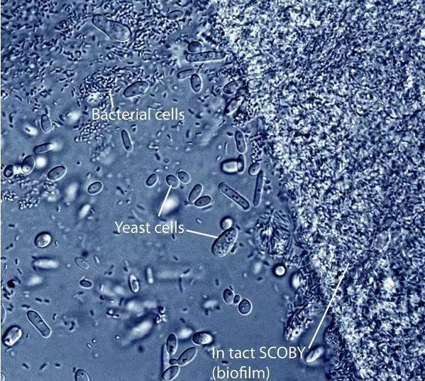
- Kombucha optical micrograph showing Gluconacetobacter bacteria and yeast cells in its semipermeable membrane.
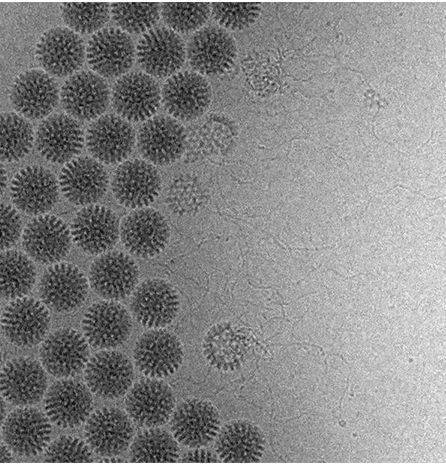
- Cryo-electron microscope image of the double-stranded RNA genome emerging from Smian rotavirus particles.
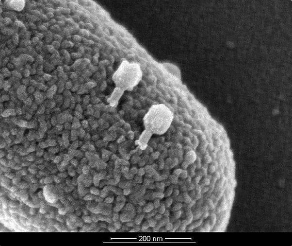
- T4 bacteriophages infecting E. coli bacteria


Electron Microscopy Today and in the Future
Transmission electron microscopes allow higher-resolution sample examination in scientific research, improving understanding of the surrounding world.
Modern electron microscopes have greater magnification and resolution than early models. Their principles, however, derive from Ernst Ruska's first prototype. The figure below shows his first electron microscope invented in 1933, as a faithful replica constructed in 1980.

Electron microscopes have exceeded optical microscopy's limits, and improved resolution enables examination of microscopic objects such as atoms. Development continues today. For example, an environmental scanning electron microscope allowing specimens to be examined in a lower-vacuum chamber is under development.

2017 Nobel Prize in Chemistry Awarded for Cryo-Electron Microscopy Research
Jacques Dubochet, Joachim Frank and Richard Henderson received the 2017 Nobel Prize in Chemistry for cryo-electron microscopy. Cryo means freezing, and this electron microscopy technique enabled high-resolution imaging of biological molecules.
Working at Cambridge University's MRC Laboratory of Molecular Biology, Prof. Richard Hendersonused electron microscopy in the 1990s to determine a protein's structure at atomic scalesuccessfully.
Working at Columbia University, Prof. Joachim Frankdeveloped an image-processing method through work between 1975-1986. Analysis of two-dimensional microscope images then allowed three-dimensional structures to be determined through this approach.
Working at the University of Lausanne, Prof. Jacques Dubochetdeveloped a method to determine molecular structures in water. Samples examined by electron microscopes are placed in very low-density environments. But when a sample is an aqueous biomolecule, water rapidly evaporates there and damages its structure. In the early 1980s, Dubochet developed a method cooling water very rapidly, solidifying biomolecules without disrupting their structures. This opened the way to determining their natural structures in aqueous environments, as in living tissues.
After years of effort, electron microscopy achieved the desired resolution for biomolecules only in 2013. Today, cryo-electron microscopy can determine structures ranging from proteins to viruses.
Sources
nobelprize.org
2. Written by Yolanda Smith.
History of the Electron Microscope.
news-medical.net - Last Updated: Jul 27, 2017
Was this article helpful?
Your feedback helps us improve our content.




Reader comments
Share your comment
Your name and comment are published only after administrator approval. Your email address is never shown to visitors.
Be the first to comment.