
What is FAPI PET? Next-generation imaging targeting cancer cells

In this article
What is FAPI PET?
FAPI PET is a next-generation form of oncological imaging that targets fibroblast activation protein (FAP) to detect cancer cells.
Fibroblast activation protein (FAP) is a molecule overproduced in cancer-associated fibroblasts. Radioactive FAP inhibitors (FAPIs) targeting these molecules are used as PET tracers, allowing tumors to be visualized almost at the cellular level.
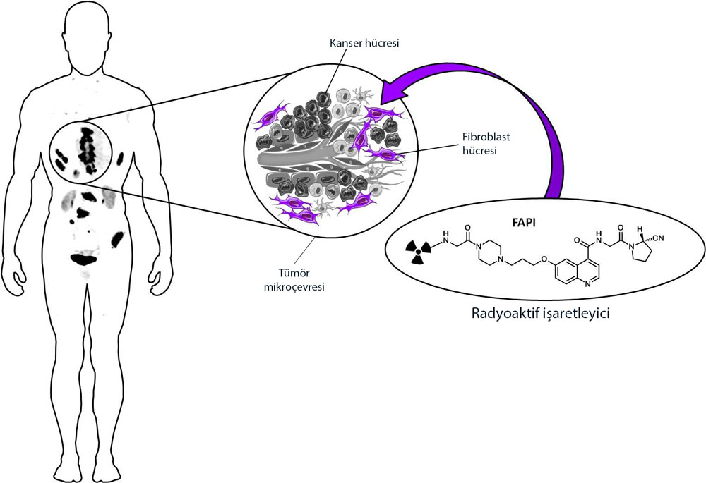
Figure explanation: After intravenous administration, FAPI, designed as a radioactive tracer, is taken up in the tumor microenvironment by the cancer-associated fibroblasts abundant there. This allows the tumor to be visualized.
Fibroblast activation protein (FAP) is abundant in some tumors. Breast, colon and pancreatic cancers, in particular, have a strong, dense tumor microenvironment, which means that 90% of the gross tumor mass may consist of stromal (noncancerous connective tissue) cells.
Although connective-tissue cells called fibroblasts are found throughout the body, cancer-associated fibroblasts occur in the tumor microenvironment and differ from normal fibroblasts by secreting FAP. Based on this scientific discovery, FAP inhibitors (FAPIs) have already been developed as cancer drugs.
This FAP-specific inhibitor is a quinoline-based molecule and has been considered highly promising as an oncological imaging tracer by targeting cancer-associated fibroblasts that produce FAP.
FAPIs are thought to have potential uses not only in imaging but also as therapeutic agents.
The most important requirement for good cancer treatment is the best possible cancer imaging
Conventional radiographs, called X-Ray in medicine, cannot visualize tumors until they reach a fairly large size, for example 3-5 cm. The introduction of computed tomography (CT) and then magnetic resonance imaging (MRI) made it possible to detect smaller tumors, for example 1 cm or even 0.5 cm. Building on this development, PET-CT and other fusion (combined) imaging methods such as PET-MR enabled us to determine both tumor size and metabolic activity: anatomical as well as functional imaging. In this form, PET scans are the most advanced oncological imaging methods.
Efforts to improve cancer imaging with PET further
PET (Positron Emission Tomography) focuses on molecular targets as a form of nuclear imaging. A PET scan targets particular molecules used by cells in the body. For example, all cells consume glucose (sugar), but because cancer cells have a faster metabolism, they use more sugar. The most commonly used FDG PET-CT uses radioactively labeled sugar molecules to visualize cells consuming sugar more intensely. Today, FDG PET-CT provides valuable information and guidance in the diagnosis and treatment of many cancers. However, FDG PET imaging also has some limitations.
FDG PET shows physiological (natural) activity uptake in organs that use glucose intensively, such as the brain, heart and liver. New molecular targets have therefore been sought to distinguish cancer cells from normal cells more effectively.
FAPI PET uses FAPI, which targets FAP produced by cancer-associated fibroblasts, instead of radioactively labeled sugar to target cancer cells.
The differences between FDG PET and FAPI PET can be seen below:

Figure explanation: FAPI PET may be particularly useful in cancers where conventional FDG PET is insufficient. For example, the images above show improved tumor visualization with FAPI PET in certain cancers in which the tumor microenvironment is prominent. As shown above, FAPI PET provides much better imaging in stomach, pancreatic, liver and ovarian cancer.
Unlike FDG PET, FAPI PET imaging does not require fasting, and the blood-sugar adjustments needed before FDG PET in people with diabetes are not required for FAPI PET. In addition, although FDG PET requires waiting 60 minutes after injection, FAPI PET imaging can begin from the tenth minute.
Not only imaging but also treatment: theranostics
The most important feature of FAPI is that it is a theranostic agent, used for both diagnosis and treatment. It can be synthesized with 68Ga for PET imaging (68Ga-FAPI PET/CT), or with radioisotopes with therapeutic properties such as 90Y (yttrium) and 177Lu (lutetium) for radionuclide treatment (90Y-FAPI, 177Lu-FAPI).
Sources
Was this article helpful?
Your feedback helps us improve our content.




Reader comments
Share your comment
Your name and comment are published only after administrator approval. Your email address is never shown to visitors.
Be the first to comment.