Ground-Glass Opacity and Subsolid Nodule in a Chest CT Report: Is It Cancer or Is It an Infection?
In this article
Ground-Glass Opacity and Subsolid Nodule in a Chest CT Report: Is It Cancer or Is It an Infection?
When a chest CT report mentions “ground-glass opacity,” “ground-glass nodule” or “subsolid nodule,” many people immediately ask: “Is it cancer?” These phrases are not diagnoses but descriptions of the image: an area where lung tissue is slightly denser, yet the lines of the vessels and airways can still be seen through it. Temporary causes such as infection, bleeding and inflammation, and also early lesions on the lung adenocarcinoma spectrum that persist, can produce the same picture. What separates them is usually not a single CT but time: in a Korean screening cohort of 50,132 adults, 30.0% of subsolid nodules found at the first screening and 78.9% of those that appeared later resolved on their own. This article explains the language of the report, temporary and persistent causes, the Fleischner and Lung-RADS follow-up recommendations, and what is done for persistent lesions. Decisions about your own report belong to the physicians who see your images.
- What does ground-glass mean? On CT, a slightly increased density in part of the lung through which vascular and bronchial structures are still visible. It is not a diagnosis but a description of the image. If round/oval and well defined, it is called a “ground-glass nodule”; if it also has a solid component, a “part-solid nodule”; together they are called “subsolid nodules.”
- Cancer or infection? One CT cannot reliably tell them apart. Temporary causes such as infection and bleeding usually shrink or disappear on follow-up CT; persistent ones may be focal fibrosis or lesions on the adenocarcinoma spectrum (AAH, AIS, MIA, invasive adenocarcinoma).
- How is it followed? Fleischner 2017 (incidental nodules): for a single pure ground-glass nodule ≥6 mm, CT at 6-12 months and, if it persists, every 2 years up to 5 years; for a single part-solid nodule ≥6 mm, CT at 3-6 months and, if it persists with a solid component <6 mm, annual CT for 5 years. If it persists and the solid component is ≥6 mm, it is considered highly suspicious.
- What raises concern? Growth in size, increasing density, a new or growing solid component. Then a tumor board, PET/CT and biopsy or surgery come into discussion.
- What it does not mean: It does not mean “they saw ground-glass, so it is cancer,” and it does not mean “it has been the same for years, so follow-up is no longer needed.” Both are inconsistent with guidelines and data.
What do the terms in the report mean?
Radiology reports can describe the same image in different words. The table below summarizes the most common phrases and what they mean in practice.
| Phrase in the report | Meaning | Why it matters |
|---|---|---|
| Ground-glass opacity (GGO) | Slightly increased lung density on CT; vessel and airway walls can still be seen | A descriptive term; on its own it does not mean cancer. It may be focal or diffuse |
| Ground-glass nodule (GGN, pure ground-glass) | A round or oval ground-glass area without a solid component | May be temporary (infection, bleeding) or persistent (early adenocarcinoma spectrum, fibrosis) |
| Part-solid (mixed) nodule | Contains a solid portion within the ground-glass that obscures vessels and airways | The size of the solid component is one of the most important risk criteria |
| Subsolid nodule | Common name for pure ground-glass and part-solid nodules | Guidelines manage these two types separately |
| Consolidation / solid component | A dense area that obscures the lines of vessels and airways | May relate to invasion; its size is reported separately |
| Lung-RADS | The ACR classification for lung cancer screening CT (categories 1-4) | Routine, incidental CTs use Fleischner; the two are not the same |
In the Fleischner guideline, nodule size is the average of the long and short axes, rounded to the nearest millimeter. A nodule is classified as part-solid only from 6 mm upward; for nodules smaller than 6 mm, follow-up is usually not required.
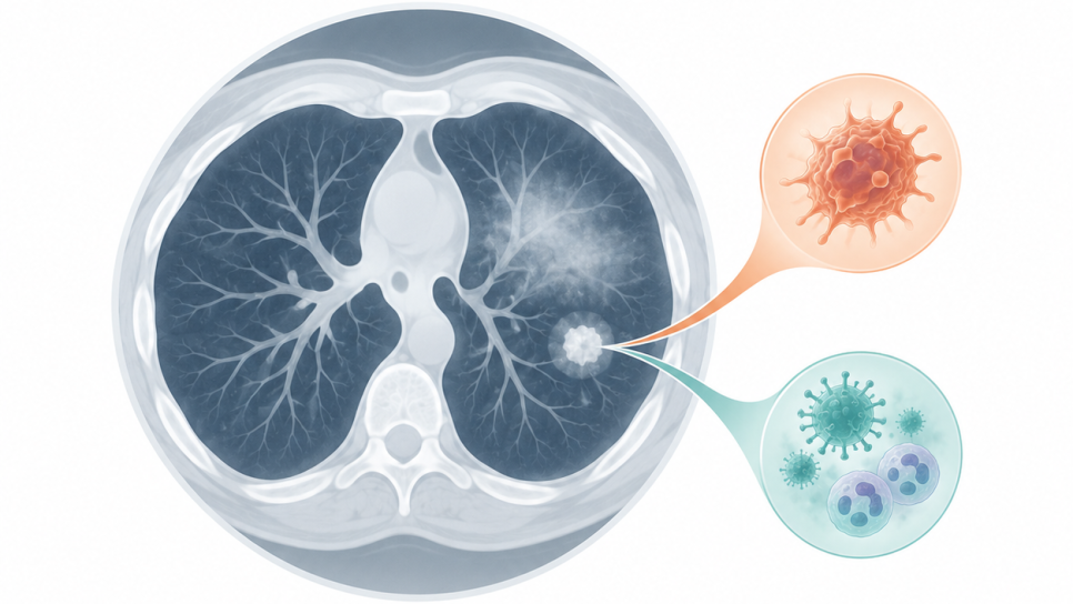
Figure 1 · Three appearances of a lung nodule
Ground-glass is a mild density that does not erase the vessel lines
Where does ground-glass come from: temporary or persistent?
The ground-glass appearance is the shared result of very different processes: partial filling of the air-filled alveoli with fluid, cells, blood or tumor cells, or thickening of the walls. For this reason it is hard to guess the cause from the image, and looking again after some time is far more informative. For incidentally found subsolid nodules, the first question is: “Does it persist?”
Figure 2 · Spontaneous resolution of subsolid nodules
Low-dose CT screening cohort of 50,132 people in Korea (Thorax 2021). The bars show only these two percentages.
In the same study, the frequency of subsolid nodules was 10.7% in never-smokers and 7.7% in ever-smokers. Of 293 nodules sampled by biopsy or surgery, 227 (77.5%) were lung cancer; because this group consists of suspicious nodules selected for sampling, it should not be read as the share of cancer among all ground-glass nodules.
- For your nodule, the follow-up CT is decisive, not the percentages. The percentages above come from a single-center Asian cohort in which healthy people were screened; resolution is much more common in nodules that appear later.
- Resolution or shrinkage should not be credited to a “course” of treatment or a medicine; temporary nodules also disappear on their own.
The cancer spectrum: from AAH to invasive adenocarcinoma
A significant share of persistent ground-glass nodules corresponds to the early and often very slowly progressing steps of lung adenocarcinoma. According to the IASLC/ATS/ERS classification and the WHO 2021 classification, the steps are these: atypical adenomatous hyperplasia (AAH) and adenocarcinoma in situ (AIS) are considered precursor lesions; minimally invasive adenocarcinoma (MIA) and invasive adenocarcinoma are separated by the presence of invasion. AIS is a tumor up to 3 cm with pure lepidic growth and no invasion; MIA is a lepidic-predominant tumor with a focus of invasion no larger than 5 mm.
Figure 3 · The adenocarcinoma spectrum and typical CT appearance
The dark center indicates the solid component
The signs considered “more invasive” on CT differ from study to study. In studies of resected pure ground-glass nodules, diameter thresholds for separating invasion were reported between 6.5 mm and 16.4 mm; that is, there is no exact line above which one can say “it is cancer.” In one study, about 10% of pure ground-glass nodules smaller than 10 mm also had foci of invasion. Imaging features linked with invasion include larger diameter and volume, an air bronchogram, lobulated or spiculated margins, and dilated vessels passing through the lesion. These are operated, selected series and cannot be applied as a rule to each patient.
Growth is usually very slow. That is why follow-up periods are long; but the reverse is also true: cases have been reported in which a ground-glass nodule stable for 5 years grew at 10-year follow-up. This shows that the 5-year limit in the guideline may not always be enough for an individual; the follow-up plan should be reviewed with your physician.
Follow-up guidelines: Fleischner 2017
The Fleischner Society’s 2017 recommendations apply to incidentally detected nodules in adults aged 35 and over. Screening CTs, immunosuppressed patients and patients with a known primary cancer are outside the scope. The table below summarizes the recommendations for subsolid nodules.
| Finding | Suggested approach | Note |
|---|---|---|
| Single pure ground-glass nodule <6 mm | No routine follow-up | With suspicious morphology, CT at years 2 and 4 may be considered; with growth or a new solid component, resection is considered |
| Single pure ground-glass nodule ≥6 mm | CT at 6-12 months to check persistence; if persistent, CT every 2 years up to 5 years | In practice often expressed as checks at around years 1, 3 and 5 |
| Single part-solid nodule <6 mm | No routine follow-up | A nodule can be classified as part-solid only from 6 mm upward |
| Single part-solid nodule ≥6 mm | CT at 3-6 months to check persistence; if unchanged and the solid component is <6 mm, annual CT for 5 years | If it persists and the solid component is ≥6 mm, it is considered highly suspicious |
| Multiple subsolid nodules <6 mm | CT at 3-6 months; if stable, CT at years 2 and 4 is considered | Multiple pure ground-glass nodules <6 mm are usually benign; in selected high-risk patients, follow-up at years 2 and 4 is considered |
| Multiple subsolid nodules ≥6 mm | CT at 3-6 months; further management is set by the most suspicious nodule | With multiple nodules, temporary (infectious, inflammatory) processes are considered more often |
The table was compiled from the summary table of the Fleischner Society 2017 guideline. Recommendations from other bodies such as NCCN, CHEST, ACR and AATS may differ in size, solid component and timing; that is, a slightly different interval recommended at two centers is not a contradiction.
Figure 4 · Typical follow-up course for a single pure ground-glass nodule ≥6 mm
Fleischner 2017; timings are approximate and vary by person
If growth, increased density or a new solid component appears, the plan changes without waiting: short-interval CT, tumor board, tissue diagnosis or surgery.
What does Lung-RADS (screening CT) say?
On low-dose CTs taken within lung cancer screening, Lung-RADS is used instead of Fleischner. According to the 2022 version (v2022), the categories for subsolid nodules and their approximate probabilities of malignancy are as follows.
| Category | Subsolid finding | Approx. malignancy | Suggested approach |
|---|---|---|---|
| 2 (benign) | Pure ground-glass <30 mm (or ≥30 mm that is stable or slow-growing); part-solid with total diameter <6 mm | <1% | Annual low-dose CT at 12 months |
| 3 (probably benign) | Pure ground-glass ≥30 mm (at baseline or new); part-solid with total diameter ≥6 mm and solid component <6 mm | 1-2% | Low-dose CT at 6 months |
| 4A (suspicious) | Part-solid with solid component ≥6 mm and <8 mm; new or growing solid component <4 mm | 5-15% | Low-dose CT at 3 months; PET/CT may be considered |
| 4B (very suspicious) | Part-solid with solid component ≥8 mm; new or growing solid component ≥4 mm | >15% | Diagnostic chest CT, PET/CT and/or tissue sampling |
In Lung-RADS, growth means an increase of more than 1.5 mm in mean diameter within 12 months. A ground-glass nodule that doubles in size within a year is a feature that raises the category. Category numbers and probabilities were compiled from the search summary of the ACR document and secondary sources; for exact categorization, see the ACR version named in your report.
What is more reassuring, and what draws more attention?
What is done if it persists and is suspicious?
A persistent, suspicious subsolid nodule is evaluated in a tumor board of pulmonologists, thoracic surgeons, radiologists and medical oncologists. Because the metabolic activity of ground-glass nodules is low, the contribution of PET/CT is limited; Lung-RADS lists PET/CT among the options for nodules with a solid component of 8 mm or more. In choosing between biopsy, surgical removal and follow-up, the location of the nodule, the patient’s lung function and preferences are decisive.
If surgery is needed, operations that remove only part of the lung rather than a whole lobe have been shown to give good results in small, ground-glass-dominant tumors. Two Japanese studies support this:
| Study | Patients | Operation | Result |
|---|---|---|---|
| JCOG0804/WJOG4507L (single-arm, confirmatory phase 3) | Peripheral, ≤2 cm, consolidation/tumor ratio ≤0.25; 333 enrolled | Sublobar resection in 314 patients (258 wedge, 56 segmentectomy) | 5-year relapse-free survival 99.7% (90% CI 98.3-99.9); no local relapse; grade ≥3 complications in 17 patients (5.4%) |
| JCOG1211 (single-arm, confirmatory phase 3) | Ground-glass-dominant, ≤3 cm including ground-glass; 396 enrolled | Segmentectomy in 357 patients | At median follow-up of 5.4 years, 5-year relapse-free survival 98.0% (95% CI 95.9-99.1); grade 3-4 early complications in 7 patients (2%); no grade 5 |
Both studies were conducted in Japan, in selected patients and without a comparison arm; the results do not apply directly to every patient. Still, they show that surgery gives good results in early ground-glass-dominant tumors, and that when the operation is not urgent, the option of follow-up can also be discussed.
- A study published in February 2026 in Radiology: Cardiothoracic Imaging examined 1,724 people with a dominant ground-glass nodule in a CT lung cancer screening program. Lung cancer was diagnosed in 114 people; 70 (61%) arose from the dominant ground-glass nodule, and 66 of these (94%) were stage 0 or I. All 10 lung-cancer-related losses of life were from solid cancers unrelated to the ground-glass nodule.
- In people with a ground-glass nodule, the likelihood of a cancer unrelated to the nodule was also higher than in those with Lung-RADS 1 (P=.007).
- Guidelines are consensus texts: Fleischner 2017 is expert opinion without formal grading of evidence; there are differences between bodies.
- Data rest on selected groups: surgical series represent only operated patients, and screening cohorts represent particular populations.
- Percentages do not apply to an individual: resolution and cancer proportions vary with population, screening and nodule type.
- This article is general information: decisions about your own report belong to the physicians who see your images.
- Which system is named in the report? “Fleischner” is used for incidental nodules and “Lung-RADS” for screening CT. Turkish teaching materials also present the Fleischner 2017 table in the same way (ASYOD presentation).
- Bring your old CTs: To assess growth, previous chest CTs (a CD or PACS access) are often the most valuable information; thin-section images are preferred.
- Who evaluates? Specialists in pulmonology, thoracic surgery, radiology and medical oncology decide together.
- A national nodule follow-up guideline or lung cancer screening program has not been confirmed in this article; clarify the follow-up plan with your treating physician.
- If a cancer diagnosis is confirmed, the decision about molecular testing rests with your physician (what is next-generation sequencing (NGS)?, in Turkish).
Questions you can ask your doctor
- Is my nodule pure ground-glass or part-solid; what is its diameter and, if present, the size of the solid component in mm?
- Which system (Fleischner, Lung-RADS) was the report based on, and when is the first follow-up CT?
- Has it been compared with my old CTs; is there growth or increased density?
- If I recently had an infection, should the follow-up CT be done sooner?
- For how many years in total will I be followed; at what finding would a tumor board or surgery come into discussion?
Common misconceptions
- “If ground-glass is seen, it is cancer.” No. Temporary causes are very common; in the Korean screening data, 78.9% of new subsolid nodules resolved on their own.
- “It has been the same for years, so follow-up is no longer needed.” No. Ground-glass nodules that were stable for 5 years and then grew have been reported; decide when to end follow-up together with your physician.
- “PET/CT gives a definite answer for every nodule.” No. The metabolic activity of ground-glass nodules is low, and the value of PET/CT is limited.
- “The nodule disappeared, so the medicine/course worked.” No. Temporary nodules also disappear on their own; do not draw a cause-and-effect link.
Frequently Asked Questions
- Is ground-glass opacity cancer?
- No, not by itself. Ground-glass is seen with temporary causes such as infection, bleeding and inflammation, and also with persistent lesions (fibrosis or lesions on the adenocarcinoma spectrum). A follow-up CT shows whether it persists.
- How can infection be told apart from cancer?
- One CT cannot reliably tell them apart. Infection and bleeding usually shrink or disappear on follow-up; persistence, growth, increased density or a new solid component point more toward the cancer spectrum. A history of recent infection helps the physician.
- How often is a ground-glass nodule followed?
- According to Fleischner 2017, for a single pure ground-glass nodule ≥6 mm, CT at 6-12 months and, if it persists, every 2 years up to 5 years is recommended; for a single part-solid nodule ≥6 mm, the first check is at 3-6 months. On screening CTs, Lung-RADS intervals are used. Your physician sets your own plan.
- Does a ground-glass nodule need surgery?
- Not always. For persistent and suspicious nodules that grow or develop a solid component, the tumor board discusses surgery. In Japanese studies of small ground-glass-dominant tumors, 5-year relapse-free survival of 98-99.7% was reported after sublobar surgery; these results apply to selected patients.
- What does having several ground-glass nodules mean?
- With multiple subsolid nodules, temporary infectious or inflammatory processes are considered more often; multiple pure ground-glass nodules smaller than 6 mm are usually benign. For those 6 mm or larger, CT is repeated at 3-6 months and management is set by the most suspicious nodule.
- Are ground-glass nodules seen in non-smokers too?
- Yes. In the Korean screening cohort, the frequency of subsolid nodules was higher in never-smokers (10.7%) than in ever-smokers (7.7%).
Glossary
- Ground-glass opacity (GGO)
- Slightly increased lung density on CT; vessel and airway structures remain visible.
- Pure ground-glass nodule (pGGN)
- A nodule without a solid component.
- Part-solid nodule
- A nodule that contains a solid component within the ground-glass.
- Subsolid nodule
- Common name for pure ground-glass and part-solid nodules.
- Solid component
- The dense part inside a nodule that obscures vessels and airways; often corresponds to the area of invasion.
- AAH (atypical adenomatous hyperplasia)
- A precursor lesion of adenocarcinoma; usually smaller than 1 cm and pure ground-glass in appearance.
- AIS (adenocarcinoma in situ)
- Adenocarcinoma up to 3 cm with purely lepidic growth and no invasion.
- MIA (minimally invasive adenocarcinoma)
- A small lepidic-predominant adenocarcinoma with an invasion area of no more than 5 mm.
- Lepidic growth
- Spread of tumor cells along the alveolar walls without destroying the existing structure.
- Fleischner guideline
- An international set of recommendations for following incidentally detected lung nodules (2017).
- Lung-RADS
- The ACR reporting system that classifies lung cancer screening CTs.
- Sublobar resection
- Removal of part of a lung lobe (wedge or segment) rather than the whole lobe.
- Relapse-free survival (RFS)
- The proportion or length of time patients remain free of relapse after treatment.
DROZDOGAN Academy Commentary
A ground-glass finding is not an “alarm signal” but “the start of follow-up”; time and comparison images supply the key information.
⚡ Why it matters
With the spread of chest CT, ground-glass findings are reported very often and can cause patients needless worry or, conversely, needless reassurance. The right question is not “is it cancer?” but “does it persist, does it change?”
✅ Strengths
The guidelines (Fleischner, Lung-RADS) are clear and usable; the frequency of temporary causes and the follow-up CT spare many patients unnecessary intervention; the results of sublobar surgery in small ground-glass-dominant tumors are very good.
⚠️ Limits
Guidelines are expert consensus and differ between bodies. The percentages come from single-center, predominantly Asian cohorts. One CT cannot reliably separate the causes; the 5-year limit may not be enough for everyone.
🩺 In practice
Take the report with your old CTs to a pulmonologist, thoracic surgeon or radiologist; get the measurements (diameter, solid component) and the follow-up plan in writing; do not miss the follow-up dates.
❓ Open questions
Which pure ground-glass nodules will really progress? In whom should follow-up extend beyond 5 years? Will artificial intelligence and radiomics be able to reduce the follow-up burden?
Conclusion: Ground-glass opacity is not a diagnosis but a follow-up question. The decision is made by observing persistence, growth, density and the solid component; make it together with the physicians who see your images, with timely follow-up checks.
- This article was prepared in November 2026.
- The Fleischner 2017 recommendations were checked against the guideline’s summary table (NYGH) and secondary summaries; the full text of the guideline was not examined directly here. The Lung-RADS v2022 thresholds and probabilities were taken from the search summary of the ACR document and secondary sources (Radiopaedia, radiogyan); small differences between the two sources were corrected according to the ACR document.
- The Thorax 2021 (Korea) figures come from the article abstract; those from Hammer et al. (2026) from the search summary of the PubMed record and publisher page; and the JCOG0804 and JCOG1211 figures from published abstracts. The full texts were not examined directly here.
- The figures are simplified and are not real CT images. The Academy commentary is our own.
- This article is not medical advice; make decisions about your health together with your physician.
Sources
- MacMahon H, Naidich DP, Goo JM, et al. Guidelines for Management of Incidental Pulmonary Nodules Detected on CT Images: From the Fleischner Society 2017. Radiology. 2017;284(1):228-243. doi:10.1148/radiol.2017161659. pubs.rsna.org
- Fleischner Society 2017 Guidelines, summary table (NYGH). nygh.on.ca
- American College of Radiology. Lung-RADS v2022. November 2022. acr.org
- Kim YW, Kwon BS, Lim SY, et al. Lung cancer probability and clinical outcomes of baseline and new subsolid nodules detected on low-dose CT screening. Thorax. 2021;76:980-988. pmc.ncbi.nlm.nih.gov
- Hammer MM, et al. Clinical Outcomes of Ground-Glass Nodules Detected in a CT Lung Cancer Screening Program. Radiol Cardiothorac Imaging. 2026;8(1):e250293. doi:10.1148/ryct.250293. pubmed.ncbi.nlm.nih.gov
- Truong MT, Ko JP, Rossi SE, et al. Update in the Evaluation of the Solitary Pulmonary Nodule. RadioGraphics. 2014;34(6):1658-1679. doi:10.1148/rg.346130092. pubs.rsna.org
- Milanese G, Sverzellati N, Pastorino U, Silva M. Adenocarcinoma in pure ground glass nodules: histological evidence of invasion and open debate on optimal management. J Thorac Dis. 2017;9(9):2862-2867. doi:10.21037/jtd.2017.08.120. pmc.ncbi.nlm.nih.gov
- Tang ER, Schreiner AM, Pua BB. Advances in lung adenocarcinoma classification: a summary of the new international multidisciplinary classification system (IASLC/ATS/ERS). J Thorac Dis. 2014 (PMID 25349701). pmc.ncbi.nlm.nih.gov
- Kobayashi Y, Mitsudomi T. Ground glass nodules with 5 years' stability can grow after 10-year follow-up: do genetic features determine the fate? (PMID 32038929). pmc.ncbi.nlm.nih.gov
- Suzuki K, Watanabe SI, Wakabayashi M, et al. A single-arm study of sublobar resection for ground-glass opacity dominant peripheral lung cancer (JCOG0804/WJOG4507L). J Thorac Cardiovasc Surg. 2022;163(1):289-301.e2. pubmed.ncbi.nlm.nih.gov
- Aokage K, Suzuki K, Saji H, et al. Segmentectomy for ground-glass-dominant lung cancer with a tumour diameter of 3 cm or less including ground-glass opacity (JCOG1211): a multicentre, single-arm, confirmatory, phase 3 trial. Lancet Respir Med. 2023;11(6):540-549. doi:10.1016/S2213-2600(23)00041-3. pubmed.ncbi.nlm.nih.gov
- Patel M, Bade B. Subsolid pulmonary nodules: From guidelines to real-world practice. CHEST Physician. chestphysician.org
- ASYOD. Toraks BT klinik yorumlama sunumu (2018). asyod.org
Was this article helpful?
Your feedback helps us improve our content.