The Non-Enhancing Tumor: How Does Amino Acid PET Reveal the Hidden Borders of Glioblastoma?
In this article
The Non-Enhancing Tumor: How Does Amino Acid PET Reveal the Hidden Borders of Glioblastoma?
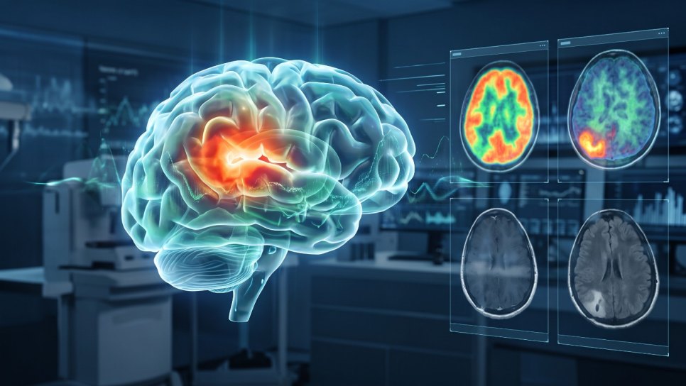
Surgery and radiotherapy for glioblastoma are planned largely on contrast-enhanced MRI. But this tumor grows by infiltrating the surrounding brain, and a significant share of the infiltrating cells do not take up contrast on MRI. In a prospective, biopsy-validated study published in Nature Communications on November 6, 2026, 472 tissue samples from 61 patients with IDH-wildtype glioblastoma were analyzed. FET-PET detected tumor in 91.2% of the tumor-containing samples, and contrast-enhanced MRI in 52.6%. Tumor was present in 98.3% of the samples taken from areas that did not enhance but lit up on FET-PET. In this article we explain what FET-PET is, why the results matter, and why it is still too early to say “FET-PET for every patient.” You can find the basics of glioblastoma in our glioblastoma guide (in Turkish).
- What was found? In 472 pathology-confirmed biopsies, FET-PET's sensitivity for showing tumor was 91.2% versus 52.6% for contrast-enhanced MRI (an absolute difference of about 39 percentage points).
- Key finding: 98.3% of tissue that did not enhance but lit up on FET-PET was tumor. In other words, some areas that MRI shows as “clean” actually contain tumor.
- Limits: A single-center study; the 472 samples came from 61 patients, and samples from the same patient are not independent of each other. Imaging has not been shown to prolong survival.
- In practice: FET-PET can complement MRI in choosing the biopsy target and assessing how far the tumor extends. The randomized GLIAA trial, which showed no benefit for radiotherapy planning in recurrent disease, calls for cautious interpretation of the results.
Why does this matter?
Glioblastoma is the most common and fastest-progressing primary brain tumor in adults. Standard treatment consists of as extensive a surgery as can be done safely, followed by radiotherapy together with temozolomide (new treatments for IDH-wildtype glioblastoma [in Turkish]). Both the area the surgeon removes and the radiotherapy target are drawn largely on the bright ring seen on contrast-enhanced MRI. Yet glioblastoma cells infiltrate beyond this ring into seemingly normal brain tissue. These invisible extensions explain why the tumor so often recurs right at the edge of the surgical cavity.
The FLAIR sequence on MRI shows a wider area; however, because this area contains edema as well as tumor cells, it cannot tell the two apart (brain edema [in Turkish]). A method that shows the true borders of the tumor more accurately could directly affect where a biopsy sample is taken, how surgery is planned and how the radiotherapy field is defined. This is why amino acid PET methods such as FET-PET have long been investigated.
What is FET-PET?
FET-PET is a PET scan in which FET, an amino acid labeled with fluorine-18 (O-(2-[18F]fluoroethyl)-L-tyrosine), is given intravenously. Because glioma cells overproduce amino acid transporters, they take up far more FET than normal brain tissue. As a result, tumor areas become visible even when the blood–brain barrier is intact.
The most commonly used type of PET in cancer is PET-CT with the sugar-like FDG; however, because normal brain tissue also uses a great deal of sugar, FDG falls short of showing the borders of brain tumors (What is PET-CT? [in Turkish]). Normal brain tissue, on the other hand, takes up amino acids only to a limited extent. This is why the difference between tumor and normal tissue is much clearer with amino acid tracers such as FET. FDOPA and methionine PET are also used for a similar purpose.
Figure 1 · The same tumor, three different images
What do contrast-enhanced MRI, FLAIR and FET-PET show?
Why does MRI miss some tumor areas?
The blood vessels of the brain are surrounded by a tight wall called the blood–brain barrier. The gadolinium-containing contrast agent used in MRI cannot cross this wall; it leaks into the tissue and appears bright only where the tumor has disrupted the vessel structure. The dense, fast-growing center of a glioblastoma breaks down this barrier; however, tumor cells that infiltrate the surrounding tissue one by one usually do not. These cells are invisible on contrast-enhanced MRI. FET, unlike the contrast agent, is taken directly into cells by amino acid transporters; so it can show tumor cells whether or not the barrier is intact.
Figure 2 · The blood–brain barrier and two different tracers
The contrast agent depends on leakage, FET depends on transporters
How was the study done?
The study was carried out prospectively at a single center in Germany, by the neurosurgery and nuclear medicine teams of Ludwig Maximilian University (LMU) of Munich. A total of 61 patients with IDH-wildtype glioblastoma were included; 44 of them were newly diagnosed and 17 were patients with recurrent disease for whom surgery or biopsy was planned (IDH and molecular diagnosis [in Turkish]). Before surgery, every patient had both FET-PET and contrast-enhanced MRI. During surgery or stereotactic biopsy, a total of 472 tissue samples were taken from points matched in location to the images. A pathologist decided under the microscope whether each sample contained tumor; the performance of the images was calculated against this “gold standard” (what is a biopsy? [in Turkish]).
Figure 3 · Where were the biopsies taken?
Four areas by contrast and FET status
What was found?
Of the tumor-containing samples, 91.2% were positive on FET-PET, whereas contrast-enhanced MRI showed contrast uptake in only 52.6% of the same samples. The absolute difference is about 39 percentage points; in relative terms, FET-PET captured tumor tissue about 1.7 times more often. The most striking finding, however, was in the non-enhancing areas: when FET-PET was positive in these areas, 98.3% of the samples taken contained tumor. In other words, tissue where FET-PET said “there is tumor here” but MRI looked normal was almost always truly tumor.
Which method showed the tumor tissue?
Sensitivity in biopsy samples confirmed by pathology to contain tumor; 61 patients with IDH-wildtype glioblastoma, 472 samples.
The absolute difference is about 39 percentage points; in relative terms, FET-PET showed tumor-containing samples about 1.7 times more often. Sensitivity was calculated at the sample level; samples from the same patient are not independent of each other.
According to the abstract the researchers presented at the ASCO congress in May 2026, the advantage of FET-PET was preserved in a similar way in newly diagnosed and recurrent patients, in the surgery and biopsy groups, and in both the dense and the infiltrating parts of the tumor. The same abstract reported FET-PET's rate of correctly showing non-tumor tissue as negative (specificity) as 82.5%, versus 33.3% for contrast-enhanced MRI, and the sensitivity of FLAIR as 85.3%. Although FLAIR's sensitivity is high, this sequence alone is not sufficient to define the borders, because it cannot tell tumor from edema.
Figure 4 · Every 100 tumor-containing samples
How many did FET-PET and contrast-enhanced MRI show?
How should we read the results?
The findings are strong and biologically coherent; however, there are some important limits. First and most important: 472 samples are not 472 separate patients. On average about 8 samples were taken per patient. Samples taken from the same patient's tumor resemble one another; therefore, proportions calculated at the sample level can make the uncertainty at the patient level look smaller than it is. Second, the study was carried out at a single experienced center; reading FET-PET and matching images during surgery require expertise, and the results need to be reproduced in other centers. Third, the points to be biopsied were not random but chosen according to the images; this may affect sensitivity calculations (reading research news correctly [in Turkish]).
The most important limit is this: this study is a diagnostic accuracy study. It proves that FET-PET shows the tumor better; but it does not show that a wider surgery or a differently drawn radiotherapy field based on this information makes patients live longer or better. A wider surgery can also increase the risk of permanent damage in regions that control functions such as speech and movement. In glioblastoma, promising laboratory or imaging findings have not always translated into patient benefit as easily as expected (laboratory findings and clinical reality [in Turkish]). That is why “seeing more tumor” and “benefiting the patient more” are not the same thing.
Figure 5 · What did this study show, and what did it not show?
Diagnostic accuracy and patient benefit
What could change in clinical practice?
Targeting the biopsy
For deep-seated tumors that cannot be removed surgically, diagnosis relies on needle (stereotactic) biopsy. If the sample is not taken from the most active part of the tumor, the diagnosis can remain incomplete or the grade of the tumor can be underestimated. The area with the highest FET-PET uptake is a good target for biopsy; this use is also included in international recommendations. The concern that a biopsy spreads the tumor is not supported by science (does a biopsy spread cancer? [in Turkish]).
Surgical planning
Some centers, where it is safe, are investigating the removal of FET-PET-positive tissue beyond the contrast-enhancing area as well (“supramaximal” or “supramarginal” resection). In a small retrospective study, a large volume of FET-PET-positive tissue remaining after surgery was associated with shorter survival; but such associations do not prove that the surgery itself prolongs survival (what is a surgical margin? [in Turkish]). The benefit of extensive surgery has to be balanced against preserving functions such as speech, movement and vision; loss of function after surgery directly affects quality of life and adherence to later treatments (rehabilitation in brain tumors [in Turkish]).
Radiotherapy planning
Although drawing the radiotherapy target with FET-PET seems logical, this idea was tested in a randomized trial and the expected result was not obtained. In the GLIAA trial, conducted at 15 centers in Germany, 200 patients with recurrent glioblastoma were assigned to groups whose re-irradiation was planned with FET-PET or with contrast-enhanced MRI. Median progression-free survival was 4.0 months in the FET-PET group and 4.9 months in the MRI group; the difference was not significant. Late serious side effects were less frequent in the FET-PET group (10% vs. 19%), but this was a secondary finding (radiotherapy and radiosurgery [in Turkish]).
Does a better image mean longer life?
GLIAA phase 3: in recurrent glioblastoma, the re-irradiation field was drawn with FET-PET or contrast-enhanced MRI; median progression-free survival, months.
The difference is not statistically significant (hazard ratio 1.14; 95% CI 0.85–1.52). Late serious side effects were reported in 10% of the FET-PET arm and 19% of the MRI arm. This trial tested only radiotherapy planning in recurrent disease.
The result of GLIAA is not directly applicable to the first radiotherapy in newly diagnosed glioblastoma, because the trial was conducted only in recurrent disease and with short-course re-irradiation. But it has an important lesson: an imaging method showing the tumor better does not by itself mean that it will benefit the patient. This benefit must be demonstrated separately in randomized trials.
Figure 6 · Steps of evidence
At which step is FET-PET in glioblastoma?
After treatment: recurrence or treatment effect?
One of the best-established uses of FET-PET today is telling whether a lesion that grows on MRI after treatment is true recurrence or a change caused by radiotherapy (pseudoprogression or radiation necrosis) (radiation necrosis [in Turkish]). This distinction can prevent unnecessary surgery or the premature abandonment of an effective treatment. The same question is often asked in brain metastases after radiosurgery (brain metastasis [in Turkish]). Still, no PET method is perfect; inflammation and some benign conditions can also show uptake (false-positive results on PET [in Turkish]). Because cortisone (dexamethasone) treatment can reduce contrast uptake on MRI, it is important to interpret the images together with this information (dexamethasone [in Turkish]).
Figure 7 · When is amino acid PET useful?
Main uses in international recommendations
The Situation in Türkiye
- Access: FET-PET is not a widely performed test in Türkiye. Which centers can perform it regularly and its reimbursement status by the SGK (Social Security Institution) could not be reliably verified at the time this article was prepared; for up-to-date information, consult your treatment team and nuclear medicine centers.
- Alternatives: Other amino acid PET tracers such as FDOPA or methionine, and advanced MRI techniques such as MR perfusion and MR spectroscopy, can be used in some centers to answer similar questions. Standard FDG PET-CT is generally not sufficient for assessing the borders of brain tumors.
- Decision process: The choice of imaging in glioblastoma should be discussed in a board that includes experts in neurosurgery, radiation oncology, medical oncology, neuroradiology and nuclear medicine (brain tumors [in Turkish]).
- Research: In studies of vaccines, cell therapies and new drugs in glioblastoma, accurate imaging is also important for both patient selection and response assessment (CAR-T in recurrent glioblastoma [in Turkish], B7-H3 CAR-T (in Turkish), personalized glioblastoma vaccine (in Turkish), what is a clinical trial? (in Turkish)).
Questions you can ask your physician
- Is contrast-enhanced MRI enough to assess the borders of my tumor, or would amino acid PET or advanced MRI techniques provide additional information?
- If a biopsy is planned, which imaging will be used to choose the most active area?
- How is the preservation of my functions being assessed when the extent of surgery is planned?
- If growth is seen on MRI after treatment, how will it be determined whether it is recurrence or treatment effect?
- Has my case been discussed at a multidisciplinary brain tumor board?
- “FET-PET replaces MRI.” No. MRI remains the best method for showing the anatomy of the brain and the structure of the tumor; FET-PET complements it.
- “If FET-PET shows more tumor, survival is longer.” This has not yet been shown. In the randomized GLIAA trial testing radiotherapy planning in recurrent disease, no benefit was found.
- “91% success in 472 patients.” No. The study had 61 patients; the number 472 refers to biopsy samples.
- “An area that does not enhance on MRI is clean.” Not always. Glioblastoma cells can also infiltrate non-enhancing tissue; this study confirmed that by pathology.
- “A normal PET-CT does the same job.” No. Because the sugar-like FDG is also taken up intensely by normal brain tissue, it is inadequate for showing the borders of a brain tumor (misconceptions about cancer [in Turkish]).
Frequently Asked Questions
- What is FET-PET?
- FET-PET is a PET scan that uses an amino acid called FET, labeled with fluorine-18. Because glioma cells take up this amino acid far more than normal brain tissue, tumor areas become visible even when the blood–brain barrier is intact.
- Is FET-PET better than MRI in glioblastoma?
- At capturing tumor tissue, yes: in a biopsy-validated study, FET-PET showed 91.2% of tumor-containing samples and contrast-enhanced MRI 52.6%. But it does not replace MRI; the two methods are used together.
- Does FET-PET prolong survival in glioblastoma?
- This has not yet been shown. The new study measured only diagnostic accuracy. The randomized GLIAA trial, which planned radiotherapy in recurrent glioblastoma with FET-PET, did not prolong progression-free survival compared with MRI.
- Is FET-PET performed in Türkiye?
- FET-PET is not a widely performed test in Türkiye, and the centers where it can be done and its reimbursement status could not be verified. Other amino acid PET tracers or advanced MRI techniques can be used for a similar purpose; discuss the options with your treatment team.
Glossary
- Glioblastoma
- The most common primary brain tumor in adults; it progresses rapidly and grows by infiltrating the surrounding brain tissue.
- IDH-wildtype
- Without a mutation in the IDH gene; one of the key criteria for the diagnosis of glioblastoma in the current classification.
- FET
- An amino acid tracer labeled with fluorine-18 that glioma cells take up in excess.
- Blood–brain barrier
- The tight structure surrounding the brain's blood vessels that limits the passage of many substances into brain tissue.
- Contrast-enhanced MRI
- MRI performed after a gadolinium-containing agent is given intravenously, showing areas where the barrier is disrupted as bright.
- FLAIR
- An MRI sequence that shows the tumor and the surrounding edema together as bright, but cannot tell the two apart.
- Sensitivity
- How many of the samples that truly have disease are correctly shown by the test.
- Positive predictive value
- The probability that disease is truly present when the test is positive.
- Stereotactic biopsy
- A tissue sample taken by reaching a point defined on imaging with a needle through a small hole in the skull.
- Pseudoprogression
- A temporary, treatment-related change that makes the tumor appear to grow on MRI after treatment but is not actually tumor growth.
DROZDOGAN Academy Commentary
This study clarifies with pathological evidence something we have long known in glioblastoma: the bright ring on MRI is not the whole tumor. How this knowledge will translate into patient benefit, however, is still an open question.
⚡ Why it matters
The prospective design and the pathological confirmation of every imaging finding provide one of the strongest pieces of evidence that amino acid PET can show non-enhancing tumor. An accuracy of 98.3% in areas that did not enhance but were FET-PET positive is clinically very meaningful.
✅ Strengths
Prospectively collected data; many location-matched biopsies; inclusion of only IDH-wildtype glioblastoma patients; consistent results in both newly diagnosed and recurrent patients.
⚠️ Limitations
Single center; sample-level analysis and many samples from the same patient; biopsy points chosen according to the images; no data on survival, loss of function or treatment decisions.
🩺 In practice
In centers where it is available, we find amino acid PET valuable especially when choosing a biopsy target and when distinguishing recurrence from treatment effect after therapy. For decisions to widen the surgical or radiotherapy field, we recommend weighing this information together with the risk of loss of function, by board decision.
❓ Open questions
Does wider surgery guided by FET-PET improve survival and quality of life? Will FET-PET radiotherapy planning in newly diagnosed glioblastoma give a different result from GLIAA? Will the findings be reproduced in multicenter studies?
Conclusion: FET-PET can reliably show the parts of glioblastoma that are invisible on MRI. This is an important step toward more accurate biopsy and better planning; but until its benefit to patients is shown in randomized trials, FET-PET should remain a tool that complements MRI.
- This article was prepared in November 2026.
- Sensitivity and positive predictive value are values taken from the Nature Communications article. Because the full text of the article could not be accessed, specificity and the sensitivity of FLAIR were taken from the ASCO 2026 congress abstract of the same research group; in this abstract the sensitivity of contrast-enhanced MRI was reported as 55.7%. Values in the published article may differ.
- The centers where FET-PET is performed in Türkiye and its reimbursement status could not be verified.
- The illustrations are simplified. The Academy commentary is our own.
- This article is not medical advice; make your health decisions together with your physician.
References
- [18F]FET PET enables histopathology-validated detection of non-contrast-enhancing glioblastoma. Nat Commun 2026. doi:10.1038/s41467-026-78980-1. nature.com
- Stuerzl R, Albert NL, et al. Histopathology-verified comparison of FET PET and contrast-enhanced MRI for tumor visualization in glioblastoma. J Clin Oncol 2026;44(16_suppl):2049. doi:10.1200/JCO.2026.44.16_suppl.2049. abstract
- GLIAA/NOA-10 ARO 2013-01 study group. O-(2-[18F]fluoroethyl)-L-tyrosine PET-guided versus contrast-enhanced T1-weighted MRI-guided re-irradiation in patients with recurrent glioblastoma. Lancet Oncol 2026. doi:10.1016/S1470-2045(25)00642-4. abstract
- Müther M, et al. 5-Aminolevulinic acid fluorescence-guided resection of 18F-FET-PET positive tumor beyond gadolinium enhancing tumor improves survival in glioblastoma. Neurosurgery 2019;85:E1020-E1029. pmc.ncbi.nlm.nih.gov
- Albert NL, et al. PET-based response assessment criteria for diffuse gliomas (PET RANO 1.0): a report of the RANO group. Lancet Oncol 2024;25:e29-e41. doi.org
- Albert NL, et al. Response Assessment in Neuro-Oncology working group and European Association for Neuro-Oncology recommendations for the clinical use of PET imaging in gliomas. Neuro Oncol 2016;18:1199-1208. doi.org
- Law I, et al. Joint EANM/EANO/RANO practice guidelines/SNMMI procedure standards for imaging of gliomas using PET with radiolabelled amino acids and [18F]FDG: version 1.0. Eur J Nucl Med Mol Imaging 2019;46:540-557. doi.org
Was this article helpful?
Your feedback helps us improve our content.