
Inflammatory Breast Cancer (IBC): A Sneaky Mastitis Mimic, Its Symptoms and Urgent Steps
In this article
Inflammatory breast cancer (IBC) is a rare, fast-moving form of invasive breast cancer that appears as redness, warmth, swelling and an orange-peel look of the breast. Because these signs resemble an infection (mastitis) so closely, IBC is often mistaken for it; the telling clue is signs that do not clear despite antibiotics or that quickly cover at least one third of the breast. Diagnosis needs a biopsy as well as an examination, and treatment usually starts with drug therapy before surgery. This article is not for diagnosing yourself, but for knowing when to act and what to ask: if the signs are not improving or are getting worse, see a doctor, preferably a breast specialist, without delay.
Who is this article for? People with a red, swollen breast or a diagnosis of mastitis that is not clearing, patients diagnosed with IBC and their relatives, and health professionals curious about the topic. This article is not medical advice; no diagnosis should be made, no treatment decision taken and no treatment already started changed on the basis of this text. Only the physicians who follow you can assess what applies to you.
What is inflammatory breast cancer?
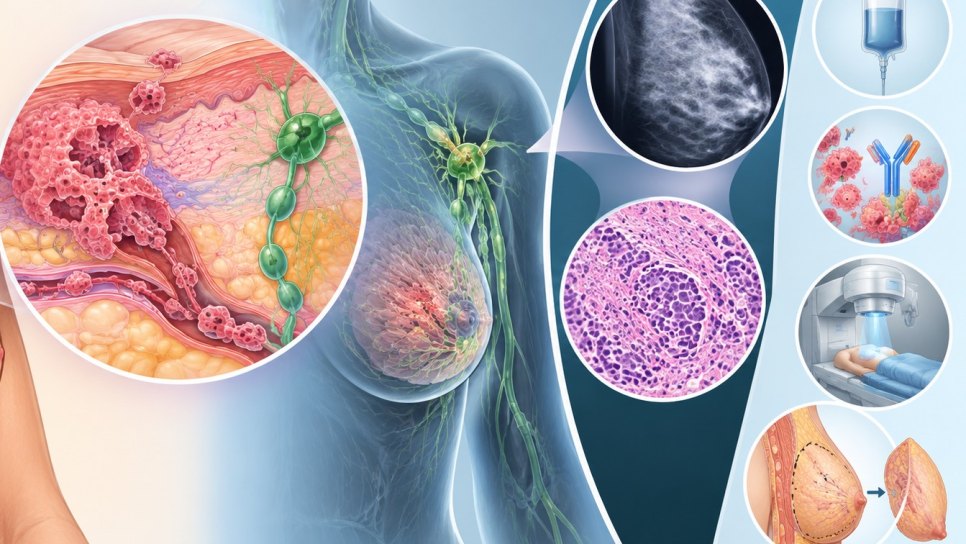
IBC is not a separate tissue (histological) type but a clinical picture: a form of invasive breast cancer that makes the breast look inflamed. In most cases the tumor is ductal, in some lobular. The redness and edema are attributed to clusters of tumor cells (tumor emboli) blocking the lymphatic vessels of the skin; that is, the name “inflammatory” comes from the appearance, not from an infection. There is often no clearly palpable lump.
| Frequency | About 1–5% of all breast cancers; in the US about 3 per 100,000 people in 1973–2015 |
| Stage | cT4d in the AJCC classification; always at least stage III at diagnosis. By definition it cannot be removed by surgery at diagnosis |
| Diagnostic criteria (AJCC 8th edition) | Diffuse redness and edema over about one third or more of the breast; less than 6 months from first symptom to diagnosis; pathological confirmation of invasive cancer |
| Pathology | Tumor emboli in the lymphatic vessels of the skin are common; however, skin findings are neither necessary nor sufficient on their own |
| Lymph node involvement | About 80% of cases have involvement of the lymph nodes |
| Distant spread at diagnosis | About 30% of patients already have disease elsewhere in the body at diagnosis (single source) |
We did not cover risk factors in this article; in the sources we reviewed we could not find shared information on them that we were able to verify.
What are the symptoms?
Symptoms usually develop over a few weeks to a few months. The American Cancer Society (ACS) lists:
- Swelling of the breast skin
- Redness covering at least one third of the breast
- Pitting or thickening of the skin that resembles orange peel
- A retracted or inverted nipple
- One breast looking larger, warmer, heavier, tender, painful or itchy compared with the other
- Swollen lymph nodes under the arm or near the collarbone
One important detail: IBC often forms no lump and may not show on a mammogram; so “nothing to feel, mammogram clear” is not reassuring on its own.
When should you seek care?
- If the breast is red and swollen, see a doctor; if mastitis is suspected, antibiotics may be the first step.
- ACS advises going back to the doctor if the signs do not improve within about a week, get worse or spread, and having additional tests for cancer.
- If there is no improvement after one or two courses of antibiotics, you are usually referred to a specialist; getting a second opinion is also advised at this point (Roswell Park).
How is it confused with mastitis?
Redness, warmth, tenderness and itching overlap with infections such as mastitis. Mastitis is very common and is most often seen during breastfeeding; in people who are not breastfeeding, causes can include a blocked milk duct, an ingrown hair or a sebaceous cyst. IBC, by contrast, is very rare. The differences that stand out in the sources are:
| Feature | Mastitis | IBC |
|---|---|---|
| Frequency | Very common | Very rare |
| Onset | Starts fairly quickly; if the infection progresses, fever and chills can develop | Described as a fast-moving cancer; signs develop over weeks to months |
| Lump | Usually none; may be felt if there is an abscess | Often none |
| Antibiotics | Usually clears with antibiotics | Patients may take antibiotics for weeks without improvement |
| Mammogram | A lump caused by mastitis does not normally show on a mammogram | Skin thickening may be seen; the mammogram can also be normal |
| Definitive distinction | Clinical assessment and response to treatment | A biopsy is needed |
Do not use this table to diagnose yourself. A trial of antibiotics can be a reasonable first step when mastitis is suspected; the real problem is waiting for longer when there is no improvement. In one review, the reported rates of IBC among people presenting with an inflamed-looking breast range from 4.5% to 50% (weighted average 16.9% across three studies); these are series from selected centers and are not a rate for the general population. The same review reports that patients most often receive an initial diagnosis of mastitis.
How is it diagnosed?
The diagnosis of IBC is first of all a clinical one and requires pathological confirmation of invasive cancer. The 2011 consensus statement of the international expert panel likewise requires the diagnosis to be supported by pathological confirmation.
- Imaging: A mammogram is usually done first; ultrasound assesses the breast and the lymph nodes under the arm; MRI is used if the mammogram and ultrasound are unclear. A mammogram may show skin and trabecular thickening, distortion and increased density; according to a review, ultrasound shows a focal abnormality in more than 90% of cases, and MRI shows diffuse edema, skin thickening and contrast enhancement, again in over 90%.
- Biopsy: A core biopsy confirms invasive cancer and determines hormone receptor and HER2 status. A punch biopsy from the affected skin may show tumor emboli in the skin; however, it does not by itself make the diagnosis, and its absence does not exclude it.
- Photographs: Photographs of the breast may be taken to document the extent of the redness.
- Pathology examination: The grade of the cancer and hormone receptor, HER2 and other gene changes are examined; this information guides the choice of treatment.
With this information the tumor is staged as “cT4d” and the treatment plan is made in a multidisciplinary board.
What are the treatment steps?
Because IBC cannot be removed by surgery at diagnosis, treatment usually starts before surgery. For stage III disease (not spread to distant organs), the sequence described in the sources is as follows; every patient's plan is different and is set in a multidisciplinary board:
- Step 1, treatment before surgery (neoadjuvant): Chemotherapy; depending on the tumor's features, drugs targeting HER2 or immunotherapy may be added. In HER2-positive tumors, antibodies targeting HER2 are used.
- Step 2, surgery: If the response to treatment is sufficient, a modified radical mastectomy and axillary lymph node dissection are performed. Breast-conserving surgery and skin-sparing mastectomy are generally not considered suitable because of how much of the skin and breast tissue the disease involves; the UK IBC working group notes that it could be considered after excellent shrinkage with multidisciplinary board review.
- Step 3, radiotherapy: After surgery, radiotherapy is given to the chest wall; according to a review, postoperative radiotherapy is almost always indicated.
- Step 4, treatment after surgery: It is determined by the tumor's molecular profile and the disease left behind; if hormone receptors are positive, hormone therapy is added. For those who do not respond to treatment before surgery, additional chemotherapy and/or radiotherapy may be planned.
In disease that has spread to distant organs (stage IV), combinations of chemotherapy, hormone therapy, targeted drugs and immunotherapy are used according to the features of the cancer.
Adherence to the recommended three-part treatment and survival
In the same National Cancer Database study, the share receiving concordant treatment fell from 51.2% in 2006–2012 to 43.9% in 2013–2019 (p<0.001). The types of non-concordance: mixed 39.7%, chemotherapy only 25.5%, radiotherapy only 24.0%, surgery only 10.8%. Simple mastectomy was found to be worse than modified radical mastectomy for the 5-year risk of loss of life (HR 1.26; 95% CI 1.08–1.46); non-concordance in surgery alone was not significantly associated with survival. The study is retrospective; the authors state that causality cannot be inferred.
Limits of the evidence. Patients who could not receive the three-part treatment may have had more advanced disease, been less robust or been steered toward a different treatment for other reasons; so these comparisons do not mean “this is how much difference it makes if you receive the treatment”. The design of a randomized trial called ConSIBreC, testing breast-conserving surgery in patients with a clinical complete response to chemotherapy before surgery, has been published (329 patients per arm, primary endpoint local recurrence 24 months after surgery); its results are not in this article's sources.
Survival and the role of subtypes
IBC does not behave as a single disease; the response to treatment before surgery and survival vary with hormone receptor (HR) and HER2 status. Among 1,061 stage III IBC patients diagnosed in 2006–2015 in the Netherlands Cancer Registry, the subtype distribution is: HR+/HER2− 42.7%, HR−/HER2− 24.3%, HR−/HER2+ 17.0%, HR+/HER2+ 16.0%. 5-year overall survival was 55.6% in all patients and 38.8% in the HR−/HER2− group; the adjusted hazard ratio for HR−/HER2− versus HR+/HER2− is 2.40 (95% CI 1.29–4.45).
Complete response rate with chemotherapy before surgery
Pathological complete response in the breast (ypT0/Tis), by subtype (%)
670 stage III IBC patients who received chemotherapy before surgery; Netherlands Cancer Registry, 2006–2015.
The overall rate is 23.2% (156/670). The combined complete response in the breast and the armpit is lower (HR−/HER2+ 36.7%; HR+/HER2− 4.0%). Bar lengths are drawn relative to the highest value.
In the same study, achieving a complete response went together with higher 5-year survival in every subgroup; but this is an association and does not show that the response alone determines survival. 5-year overall survival between patients who received the three-part treatment and those who did not is as follows:
| Subtype | Received three-part treatment | Did not |
|---|---|---|
| HR+/HER2− | 74.9% | 46.1% |
| HR+/HER2+ | 80.4% | 52.6% |
| HR−/HER2+ | 76.4% | 29.7% |
| HR−/HER2− | 47.6% | 27.8% |
Six common misunderstandings:
- “A red, swollen breast is mastitis.” Often it is, because mastitis is very common; but if there is no improvement despite antibiotics, another cause must be sought.
- “If there is no lump, there is no cancer.” No. IBC often forms no lump.
- “If the mammogram is normal, there is no problem.” No. A mammogram can be normal in IBC; diagnosis rests on clinical signs and biopsy.
- “If a skin biopsy shows no tumor emboli, IBC is excluded.” No. A skin finding is neither necessary nor sufficient on its own.
- “IBC is treated by starting with surgery.” Generally no. Treatment usually starts with drug therapy before surgery.
- “Breast-conserving surgery is always possible.” No. It is generally not considered suitable; exceptions are considered in a multidisciplinary board.
Questions you can ask your oncologist
If you are being seen for a red, swollen breast or have been diagnosed with IBC, you can ask:
- Do my findings meet the IBC criteria (extent of redness, duration, biopsy)?
- Which biopsies were done (core biopsy, skin biopsy)? What are my hormone receptor and HER2 results?
- Which imaging tests are planned to see whether the disease has spread elsewhere?
- What is my plan for treatment before surgery? If HER2 is positive, will a targeted drug be added?
- How and when will we assess the response to treatment? When will surgery be planned?
- Which surgery is recommended, and why might a breast-conserving option not be suitable?
- What is my plan for radiotherapy and treatment after surgery? Is there a clinical trial that suits me?
The situation in Türkiye. For this article we did not check how many patients in Türkiye are diagnosed late with IBC, access to multidisciplinary breast clinics, drug reimbursement, or access to clinical trials; we have drawn no assumptions on these. For how molecular tests contribute to treatment choice, see our article on next-generation sequencing (NGS) (in Turkish), and for one way to reach new treatments, our article on taking part in clinical trials (in Turkish).
DROZDOGAN Academy Commentary
IBC is a rare picture that has to be told apart: its signs resemble a common infection, but often there is no lump, antibiotics do not work, and the treatment plan starts with drugs before surgery. Using time well in diagnosis and treatment is the most consistent message in the sources.
⚡ Why it matters
Mastitis is very common and IBC very rare; so the first diagnosis that comes to mind is usually mastitis, and there is a risk of delay in diagnosing IBC (this assessment is ours). Considering additional tests without prolonged waiting for redness and swelling that do not clear is a warning that both patient and physician should keep in mind.
✅ What is strong
The diagnostic criteria (AJCC cT4d, the international expert panel) and the order “systemic treatment before surgery, then mastectomy and radiotherapy” are described consistently across several sources. Guideline-concordant three-part treatment was associated with better survival in a large series of 13,733 patients.
⚠️ What is weak
Most survival data come from retrospective registry analyses; patients who could not receive treatment may differ in other ways. The share of IBC among patients mistaken for mastitis rests on series from selected centers and spans a very wide range (4.5%–50%). There is no randomized data on the place of breast-conserving surgery in this article's sources. We could not find verified shared information on risk factors.
🩺 What it means in practice for patients and physicians
If there is new redness and swelling in the breast, taking a photograph, noting when it began and, if it does not clear with antibiotics or gets worse, seeing a breast specialist without delay are reasonable steps. Once diagnosed, knowing the subtype (HR and HER2) guides treatment, and planning in a multidisciplinary board matters.
❓ Open questions
Could breast-conserving surgery be safe after a complete response in selected patients? Which new approaches will improve survival in triple-negative IBC? What practical algorithms would reduce diagnostic delay? The answers to these questions are not in this article's sources.
Bottom line: IBC is a rare, fast-advancing breast cancer with diffuse redness and edema of the breast skin; it can look like mastitis, but it does not respond to antibiotics and is diagnosed by biopsy. Treatment usually starts with drugs before surgery, followed by mastectomy and radiotherapy. A red, swollen breast that does not clear or gets worse should not be left waiting; see a breast specialist for additional tests.
Frequently asked questions
- What is inflammatory breast cancer?
- It is a rare, fast-moving invasive breast cancer that makes the breast look red, warm and swollen. Its appearance resembles an infection; but the cause is the blocking of the lymphatic vessels of the skin by tumor cells.
- How is IBC told apart from mastitis?
- Mastitis is very common and usually clears with antibiotics; IBC is very rare, often forms no lump and does not respond to antibiotics. The definitive distinction is made by biopsy.
- How long should antibiotics be given a chance?
- We cannot give an exact number of days. ACS advises going back to the doctor if the signs do not improve within about a week or get worse; Roswell Park advises referral to a specialist and a second opinion if there is no improvement after one or two courses of antibiotics.
- Can it be IBC if the mammogram is normal?
- Yes. IBC often forms no lump and may not show on a mammogram; diagnosis rests on clinical signs and biopsy.
- How does IBC treatment begin?
- It usually begins with systemic treatment before surgery (chemotherapy; together with drugs targeting HER2 if HER2 is positive). If the response is sufficient, a modified radical mastectomy and axillary lymph node dissection are performed, followed by radiotherapy.
- Can breast-conserving surgery be done?
- It is generally not considered suitable. The UK IBC working group notes that it could be considered after excellent shrinkage with multidisciplinary board review; the results of a randomized trial on this are not in this article's sources.
- What is the survival?
- In stage III patients in the Netherlands, 5-year overall survival is 55.6%; but it varies markedly with the subtype and with whether the treatment was completed (38.8% in triple-negative). These figures are not a personal prediction.
Glossary
- Inflammatory breast cancer (IBC)
- A rare, fast-moving form of invasive breast cancer that makes the breast look inflamed.
- cT4d
- The code of inflammatory breast cancer in the AJCC classification; it means at least stage III at diagnosis.
- Orange-peel appearance
- Pitting and thickening of the skin resembling orange peel, due to disturbed lymph flow in the skin (peau d’orange).
- Tumor emboli
- Clusters of tumor cells blocking the lymphatic vessels of the skin.
- Mastitis
- Inflammation of the breast; most often seen during breastfeeding and usually clears with antibiotics.
- Neoadjuvant treatment
- Drug treatment given before surgery (such as chemotherapy).
- Modified radical mastectomy
- An operation in which the whole breast and the lymph nodes under the arm are removed.
- Pathological complete response (pCR)
- No invasive tumor left in the breast in the surgical specimen (ypT0/Tis in the study in this article).
- Hormone receptor (HR) and HER2
- Features of the tumor cells that determine the choice of treatment; they are examined in the biopsy sample.
- Core biopsy and punch biopsy
- A core biopsy takes a piece of tissue with a needle; a punch biopsy takes a small, round sample from the skin.
Transparency note. Verified: symptoms, frequency, diagnostic process and treatment sequence (American Cancer Society; Merck Manual professional version); the distinction between mastitis and IBC (Roswell Park); AJCC diagnostic criteria, imaging findings and misdiagnosis rates (the narrative review “Why diagnosing inflammatory breast cancer is hard…”); the international expert panel (Dawood et al., Ann Oncol 2011, abstract only); lymph node involvement, distant spread, “chemotherapy before surgery is the first treatment” and breast-conserving surgery (the ConSIBreC trial protocol, Front Oncol 2024; the reference to NCCN is as relayed in that paper); the UK view (Br J Cancer 2015); the national database analysis (NCDB, 2006–2019, Breast Cancer Res Treat 2025); subtype distribution, complete response and survival (Netherlands Cancer Registry, stage III IBC). Secondary relay only: the full texts of the NCCN and ESMO guidelines could not be accessed; statements about them were taken from the papers above. Not verified, and therefore not written in: detailed drug regimens in the current guideline texts; detailed treatment and survival data for stage IV disease; risk factors (age, race, weight); an exact number of days for an antibiotic trial; practice in Türkiye; overlap with existing articles on drozdogan.com (not checked). Small inconsistencies: in the abstract of the Dutch study the number of patients receiving chemotherapy before surgery is given as 679 (85.0%), while in the results section it is 670 (63.1%); we wrote the 670 used in the complete response analysis. The detailed definition of the three-part treatment and the journal in which the study was published could not be verified. Frequency figures in the sources range around 1–5%. Assessments and commentary belong to this article. This article is not medical advice; no diagnosis should be made and no treatment decision taken on the basis of this text.
References
- American Cancer Society. Inflammatory Breast Cancer. cancer.org
- Merck Manual Professional Version. Inflammatory breast cancer. merckmanuals.com
- Roswell Park Comprehensive Cancer Center. Mastitis or breast cancer? Know the difference. 2023. roswellpark.org
- Dawood S, et al. International expert panel on inflammatory breast cancer: consensus statement for standardized diagnosis and treatment. Ann Oncol. 2011;22(3):515–523. pubmed.ncbi.nlm.nih.gov
- Why diagnosing inflammatory breast cancer is hard and how to overcome the challenges: a narrative review. cco.amegroups.org
- Inflammatory breast cancer: time to standardise diagnosis, assessment and management, and for the joining of forces to facilitate effective research. Br J Cancer. 2015. nature.com
- Conserving surgery in inflammatory breast cancer after neoadjuvant chemotherapy in patients with clinical complete response: the ConSIBreC randomized controlled trial. Front Oncol. 2024. frontiersin.org
- National trends and survival outcomes associated with non-guideline-concordant treatment of inflammatory breast cancer. Breast Cancer Res Treat. 2025. link.springer.com
- Pathologic complete response and overall survival in breast cancer subtypes in stage III inflammatory breast cancer (Netherlands Cancer Registry, 2006–2015). springermedizin.de
Was this article helpful?
Your feedback helps us improve our content.



