
In Which Cancers Is FAPI PET Superior to Conventional FDG PET?

In this article
PET scans have revolutionized diagnosis and staging in oncology by revealing tumors' metabolic activity. This imaging method can detect areas of active cancer cells with high sensitivity, playing a critical role in guiding treatment and monitoring response. Used particularly to identify recurrence and metastases , PET is an important tool that has transformed oncology through its potential to improve the course of disease for cancer patients.
Fibroblast activation protein (FAPI) PET imaging is emerging as an important innovation that overcomes the limitations of conventional FDG PET/CT to detect cancers with greater sensitivity and specificity. As a protein highly expressed in cancer-associated fibroblasts (CAFs), FAPI plays a decisive role in the tumor microenvironment and tumor progression. Recent studies have shown that FAPI provides more effective tumor imaging, particularly in cancers with dense stroma (supporting connective tissue). This article examines the main cancers in which FAPI PET/CT is superior to conventional FDG PET/CT in light of scientific findings.
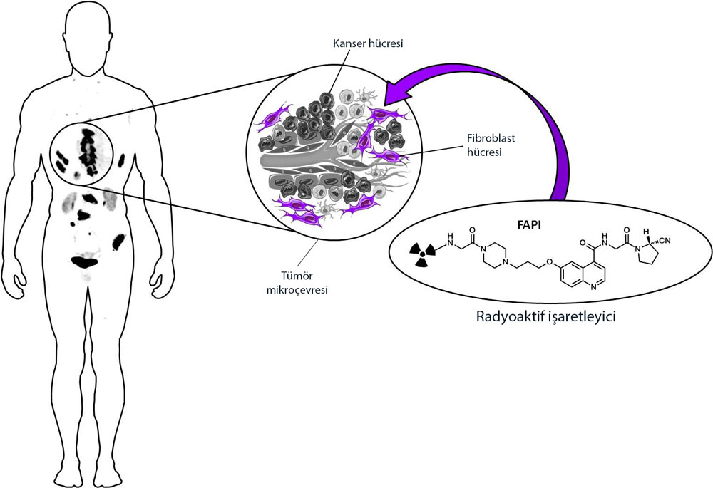
Explanation of the figure: After intravenous administration, FAPI, designed as a radioactive tracer, reaches the tumor microenvironment and is taken up by the cancer-associated fibroblasts found there in abundance. This allows the tumor to be visualized.
A New Era in Cancer Diagnosis: FAPI PET and Its Advantages
Conventional FDG PET provides effective imaging, particularly for cancers with prominent glucose metabolism. However, its low sensitivity and specificity in certain cancer types have created a need for alternatives. By providing high-quality imaging in tumors rich in cancer-associated fibroblasts (CAFs), FAPI PET has become an important option where FDG PET is limited.
CAFs form a supporting structure in the tumor microenvironment and also secrete immunosuppressive factors. FAPI's ability to bind specifically to CAFs provides greater accuracy in cancers rich in these structures.

Explanation of the figure: FAPI, a new imaging agent, is used clinically to diagnose and manage many cancers and their metastases. It has been observed to provide a higher tumor-to-background ratio than FDG, with particularly low fluctuations and variation in maximum standardized uptake value (SUVmax) in ovarian and colorectal cancers. The figure shows the tumor types in which FAPI imaging can be used.
Differences between FDG PET and FAPI PET for several cancers are shown below:

Explanation of the figure: FAPI PET may be particularly useful in cancers where conventional FDG PET is inadequate. For example, the images above show better visualization of tumors in certain cancers where the microenvironment is prominent. As shown, FAPI PET provides much better imaging in gastric, pancreatic, liver and ovarian cancers.
Liver Tumors: Hepatocellular Carcinoma (HCC) and Cholangiocarcinoma (CC)
Liver cancers are a major health problem worldwide and are known for high mortality. Primary liver tumors consist of two main histologic cancer types: hepatocellular carcinoma (HCC) and cholangiocarcinoma (CC). HCC arises from the liver's own cells and accounts for 75% of all liver cancers, while CC arises from the bile ducts and occurs at a rate of 12-15%.
Liver cancers are often diagnosed at an advanced stage, limiting the options for curative treatment. Imaging is critical for diagnosis and staging. In particular, 18F-fluorodeoxyglucose (FDG) positron emission tomography/computed tomography (PET/CT), in extrahepatic metastatic disease assessment, is widely used but has limited success in detecting primary liver cancers. Conventional FDG-PET is weak at detecting cancerous lesions within the liver.
The recently developed 68Ga-fibroblast activation protein inhibitor (FAPI) PET/CT targets fibroblast activation protein, which is highly expressed in cancer-associated fibroblasts. This provides greater sensitivity in liver tumors, particularly cancers with a dense stromal component, such as CC , improving detection.
Studies have reported sensitivity for FAPI PET/CT in detecting primary HCC of 98.5%, and for CC of 97.6% . By contrast, FDG PET/CT sensitivity was calculated at only 56.1% . The limited sensitivity of FDG PET/CT is associated with the liver's high glucose metabolism. The absence of these limitations with FAPI PET/CT offers the advantage of clearer visualization of liver tumors.
Detecting Extrahepatic Metastases
FAPI PET/CT has also shown high sensitivity in evaluating extrahepatic metastases. Studies found sensitivity of 92.2% for FAPI PET/CT in detecting extrahepatic metastatic disease, compared with 72.4% for FDG PET/CT.
In conclusion, FAPI PET/CT offers high accuracy in diagnosing both primary liver tumors and metastatic disease, emerging as a new option where FDG PET/CT falls short. However, as a new method, it requires validation in larger multicenter studies.
Pancreatic Cancers: Pancreatic Ductal Adenocarcinoma (PDAC)
Pancreatic cancer has a stroma-dominant tumor structure, making FAPI PET more effective than FDG PET for imaging this cancer. In pancreatic ductal adenocarcinoma (PDAC), FAP is highly expressed in stromal fibroblasts around the tumor. Studies show that FAPI PET provides higher tumor-to-background ratios and is more successful at detecting lymph node and distant metastases. Given PDAC's high mortality, more sensitive imaging offers a major advantage for early diagnosis and treatment.
Gastrointestinal Cancers: Gastric and Colorectal Cancers
Various studies have confirmed that FAPI PET offers greater sensitivity and clarity than FDG PET in gastric and colorectal cancers. In gastric cancer in particular, it has shown high sensitivity in diffuse and intestinal-type tumors, clearly detecting even small lesions with low glucose metabolism. This leads to a preference for FAPI PET in subtypes such as signet-ring cell carcinoma and mucosal adenocarcinoma, where FDG PET is inadequate.
In colorectal cancer, FAPI PET is reported to offer greater sensitivity than FDG PET, particularly for metastatic involvement such as lymph node and peritoneal spread. By helping define tumor margins and facilitating surgical planning, it has shown its value as a tool for improving treatment outcomes.
The Superiority of FAPI PET in Breast Cancer Staging
Accurate staging is critical for effective breast cancer treatment and patient management. A comparative analysis of FAPI PET/CT and 18F-FDG PET/CT for systemic staging of newly diagnosed breast cancer found greater staging accuracy with FAPI PET/CT. Its ability to restage approximately 20% of patients highlights its clinical effectiveness.
The widely used 18F-FDG PET/CT has certain limitations in breast cancer, including false-positive results in inflammatory breast lesions and low sensitivity in some subtypes. Its limited accuracy can result in inflammatory responses frequently being interpreted as false positives, affecting clinical decisions and leading to inappropriate treatment planning.
By targeting fibroblast activation protein, 68Ga-FAPI PET/CT shows high sensitivity for cancer-associated fibroblasts and provides more effective detection of primary tumors and metastases in breast cancer. Recent studies suggest greater sensitivity for breast cancer staging with 68Ga-FAPI PET/CT than with conventional FDG PET/CT. For example, in a study of 121 newly diagnosed breast cancer patients, FAPI PET/CT restaged 19.8% of patients, optimizing their treatment plans. Additional findings, such as bone lesions and internal mammary lymph nodes detected with FAPI PET/CT, directly affected treatment planning.
By providing accurate breast cancer staging, FAPI PET/CT contributes to personalized treatment strategies. Its use at initial staging has the potential to improve outcomes by reducing unnecessary treatments. Accordingly, using FAPI PET/CT for initial staging is expected to increase treatment effectiveness and positively affect quality of life.
Lung Cancers: Subtypes with Low Metabolic Activity
In lung cancer, FAPI PET provides better results than FDG PET, particularly in subtypes with low glucose metabolism. Conventional FDG PET is more suitable for tumors requiring high metabolic activity, whereas FAPI PET offers broader diagnostic coverage by focusing on fibroblast activation in the tumor microenvironment. This makes it superior, particularly for monitoring metastatic spread.
Head and Neck Cancers
FAPI PET/CT also offers greater diagnostic accuracy than FDG PET in head and neck cancers, providing an important advantage in detecting metastatic lymph nodes. Studies report greater sensitivity and clearer results where FDG is limited by high background activity.
Other Cancers: Sarcomas and Gliomas
FAPI PET also provides greater diagnostic accuracy than FDG PET in certain other cancers, including sarcomas and gliomas. Particularly in sarcomas with dense stroma, it offers an advantage through clearer tumor visualization.
The Role of FAPI PET in Clinical Practice and Future Directions
In conclusion, FAPI PET is an important innovation in cancer diagnosis and treatment, offering greater accuracy where conventional FDG PET is limited. It is a method with demonstrated efficacy, particularly in liver, pancreatic, gastric, colorectal, lung, and head and neck cancers, where its greater sensitivity and specificity stand out. These advantages position FAPI PET as a more broadly applicable tool in future oncologic imaging.
Testing FAPI PET in more patients and multicenter studies will enable wider clinical use. In tumors where FDG PET is inadequate or has low sensitivity, FAPI PET may contribute to more effective, personalized cancer management strategies. Its role and range of applications in oncology are therefore expected to expand.
Sources
2. Chandekar, K. R., Prashanth, A., Vinjamuri, S., & Kumar, R. (2023). FAPI PET/CT Imaging—An Updated Review. Diagnostics, 13(2018). doi:10.3390/diagnostics13122018.
3. Dong, Y., Zhou, H., Alhaskawi, A., Wang, Z., Lai, J., Yao, C., ... & Lu, H. (2023). The Superiority of Fibroblast Activation Protein Inhibitor (FAPI) PET/CT Versus FDG PET/CT in the Diagnosis of Various Malignancies. Cancers, 15(1193). doi:10.3390/cancers15041193.
4. Parida, G. K., Singhal, T., Rahman, A., & Agrawal, K. (2024). Diagnostic Performance of FAPI PET/CT vs. 18F-FDG PET/CT in Evaluation of Liver Tumors: A Systematic Review and Meta-analysis. Molecular Imaging and Radionuclide Therapy, 33, 77-89. doi:10.4274/mirt.galenos.2024.99705.
Was this article helpful?
Your feedback helps us improve our content.




Reader comments
Share your comment
Your name and comment are published only after administrator approval. Your email address is never shown to visitors.
Be the first to comment.