Invasive Micropapillary Carcinoma (Breast, Lung, Bladder): Why Does It Tend to Spread?
In this article
Invasive Micropapillary Carcinoma (Breast, Lung, Bladder): Why Does It Tend to Spread?
Many people who see the word "micropapillary" in a pathology report assume it automatically means a "worse course." The reality is more detailed: the micropapillary appearance often goes together with lymph node involvement at diagnosis and lymphovascular invasion, but how this translates into survival is not the same in the breast, lung and bladder. This article is general information; only your own doctor can say what your report means, weighing the type of disease and other findings together.
- What is micropapillary? Under the microscope, small clusters of cells lying in clear, space-like areas, without a vascular connective-tissue core in the middle. In the clusters, the surface faces outward ("reversed polarity").
- In the breast: lymph node involvement and lymphovascular invasion at diagnosis are common; however, overall survival did not differ from ordinary invasive ductal carcinoma in many analyses, while regional recurrence was found to be higher.
- In the lung: the micropapillary pattern counts as high-grade; even a small amount has been reported to be linked to a worse course.
- In the bladder: more advanced stage is seen at cystectomy; but once standard factors were taken into account, histology did not come out as an independent difference.
- Note: This appearance alone does not determine a person's course; it is interpreted together with stage, grade and other findings.
In brief, what does it tell us?
Micropapillary carcinoma is a subtype in which the cells appear under the microscope as small, hollow clusters lying in space-like areas. This appearance often goes together with lymphovascular invasion and lymph node involvement at diagnosis. That is why it is said that "the tendency to spread stands out"; but its effect on survival varies by organ and has not been shown in the same way everywhere.
| Finding that stands out at diagnosis | Survival finding | |
|---|---|---|
| Breast | Lymph node involvement and lymphovascular invasion are common | Overall survival not different in many analyses; regional recurrence higher |
| Lung | Linked to lymph node, lymphatic and pleural invasion; a grade 3 criterion | Worse disease-free and overall survival reported |
| Bladder | More advanced stage at cystectomy | Crude survival lower; no independent difference once standard factors are adjusted for |
Sources: one review for the breast, IASLC 2020 and two papers for the lung, a multicenter cystectomy series for the bladder (details below).
Figure 1 · From the microscope to the clinical question
Appearance, invasion and course are linked but are not the same thing
What does it look like under the microscope?
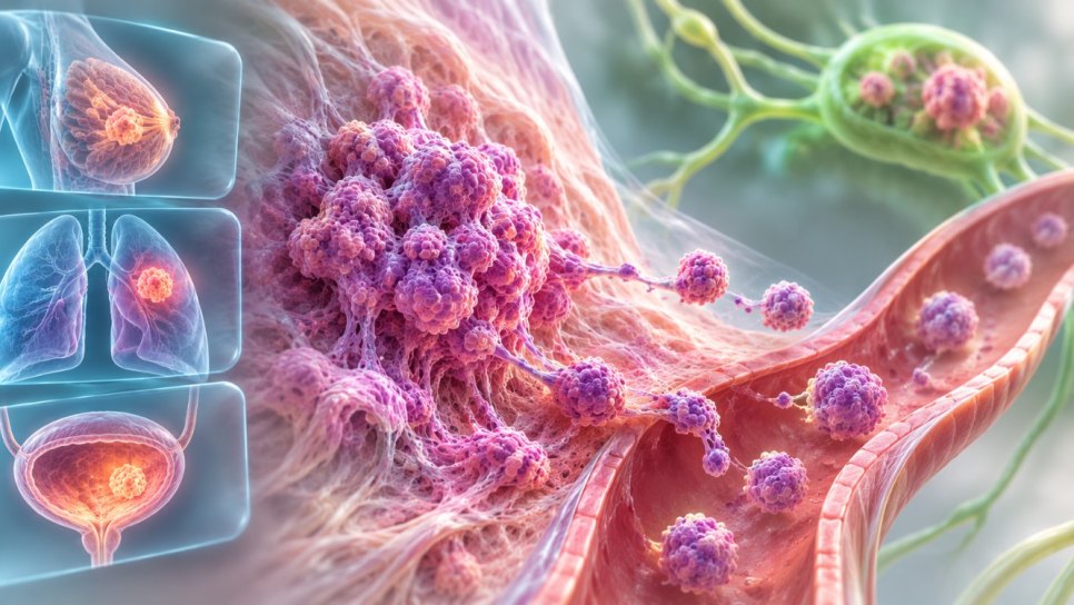
In the breast, invasive micropapillary carcinoma consists of cells arranged in hollow, pseudopapillary clusters; there is no vascular connective-tissue core (fibrovascular core) in the middle of the clusters. The lumen-facing surface of the cells faces the outside of the cluster; this is called "reversed polarity" (inside-out). MUC1/EMA staining is the best method to show this arrangement. According to the review, there is no firm guideline on what percentage of micropapillary component makes a tumor "partly" or "purely" micropapillary in the breast.
In lung adenocarcinoma, the classic appearance consists of papillary tufts without a vascular core, florets and round clusters of cells within the alveoli. Two more recently described forms exist: the "filigree" form, with thin, lace-like stacks of cells, and the "stromal" form, with clusters infiltrating the stroma within cleft-like spaces.
Why does the tendency to spread stand out?
No definite cause has been shown; what exists are findings seen together and proposed explanations.
- Lymphovascular invasion is common: in the breast, rates ranging from 14.7% to 94.7% have been reported depending on the study; in one study 77.8% in micropapillary versus 24.8% in ordinary ductal carcinoma.
- Lymph node involvement at diagnosis: 55.2% at diagnosis in a 2,660-case series in the breast; 58.1% in micropapillary-predominant tumors in the lung; 50% at cystectomy in the bladder.
- A proposed explanation: in a colorectal case report, it was suggested that the cells secrete from the surface facing the stroma, that basal MUC1 expression may disrupt adhesion to the tissue, and that basal metalloproteinase secretion may make it easier to dissect through the stroma and to invade lymphovascular spaces early. The authors present this as a suggestion, not as a proven cause.
- Mixed histology: most cases are a mixture of micropapillary and another type; this makes molecular studies and comparisons harder.
What is known in the breast?
In the breast, pure invasive micropapillary carcinoma makes up about 0.9–2% of all breast cancers; the micropapillary architecture is seen in 2–8%. Hormone receptor positivity is reported to be higher than in ordinary ductal carcinoma; in a series of 865 cases, 75.3% were luminal A, 14.8% luminal B, 4.7% HER2-enriched and 5.2% triple-negative. HER2 results are inconsistent between studies.
| Question | Finding |
|---|---|
| Overall survival | No significant difference in overall and disease-free survival in a matched analysis; in one study reported as better than ordinary ductal carcinoma (HR 0.67) |
| Regional recurrence | Higher than ordinary ductal carcinoma in two meta-analyses (OR 2.82 and 3.60) |
| A small series | In 33 micropapillary and 347 ordinary cases, lymph node involvement at diagnosis 78.2% versus 46.2%; 3-year overall survival 97% versus 94.2% (difference not significant) |
| Surgery | No evidence supports a more radical axillary approach; careful evaluation of the axilla is recommended |
| Adjuvant chemotherapy | Its effect on survival in micropapillary carcinoma has not been studied |
Most numbers are as reported in the review; the studies were done in different countries with different criteria. In the small series, the authors note that the similar outcome may have come from receiving more treatment.
What is known in the lung?
In the IASLC 2020 grading proposal, grade 1 is lepidic-predominant tumors; grade 2 is acinar- or papillary-predominant tumors with 20% or less high-grade pattern; grade 3 is any tumor with 20% or more high-grade pattern. The high-grade patterns are solid, micropapillary and complex gland patterns.
Figure 2 · Grade in lung adenocarcinoma
The predominant pattern and the percentage of high-grade pattern are used together
According to one review, micropapillary-predominant and solid-predominant tumors are among the groups with the highest likelihood of recurrence; the micropapillary pattern is linked to lymphatic and pleural invasion, lymph node involvement and advanced stage. The same article reports that in completely resected stage IB micropapillary- or solid-predominant tumors, adjuvant chemotherapy improved disease-free survival but not overall survival.
In a single-center series, 897 of 986 lung adenocarcinomas of 3 cm or smaller were found to be invasive; 344 of them (38.4%) had a non-predominant micropapillary pattern (at least 5%). Five-year disease-free survival was 67.6% versus 85.5%, and overall survival 73.5% versus 88.0%. The difference was preserved after matching as well (HR 1.862 for disease-free survival; HR 2.111 for overall survival).
| Predominant pattern | Lymph node involvement (N1 + N2) |
|---|---|
| Micropapillary | 58.1% (18 of 31 cases) |
| Solid | 50.0% |
| Acinar | 33.5% |
| Papillary | 21.4% |
| Lepidic | 10.9% |
Source: a retrospective single-center series of 986 patients (lymph node involvement 26.4% across all cases). The micropapillary-predominant group is small, so the results should be interpreted with caution.
What is known in the bladder?
In a cystectomy series of 1,497 patients from five centers, 151 patients had micropapillary urothelial carcinoma and 1,346 had urothelial carcinoma without variants. A more advanced stage was seen in the micropapillary group, but once standard clinical and pathological factors were taken into account, histology did not come out as an independent risk factor for recurrence (P = 0.27) or loss of life (P = 0.12).
| At cystectomy | No variant | Micropapillary |
|---|---|---|
| Organ-confined disease | 60% | 27% |
| Lymph node positive | 22% | 50% |
| Lymphovascular invasion | 29% | 58% |
| 5-year recurrence-free survival | 70% | 44% |
| 5-year overall survival | 61% | 38% |
Source: Mitra et al., Urologic Oncology 2019. The survival difference is clear in the crude data; in the multivariable analysis, an independent effect of histology could not be shown.
Whether guidelines should recommend radical cystectomy as first treatment for the micropapillary variant in superficial (non-muscle-invasive) bladder cancer is debated. According to a conference summary, the 2024 AUA/SUO guideline recommends repeat TUR within 4–6 weeks if bladder preservation is considered in variant histology, and says the clinician "should consider" offering radical cystectomy as initial treatment; these statements are at the level of expert opinion. The same summary stresses that the evidence in this area is of low quality and that the studies are small and retrospective. It is not certain which approach is superior; the decision is individualized.
Does a micropapillary appearance always mean a worse course?
- "Micropapillary automatically means a worse course." No. In the breast, overall survival did not differ in many analyses; in the bladder, the difference was not independent once standard factors were adjusted for.
- "It is the same in every organ." No. In the lung, the micropapillary pattern counts as high-grade in grading and has been reported to be linked to worse survival; the picture is different in the breast and bladder.
- "A small micropapillary component does not matter." No. In the lung, even a component of a few percent can carry information; that is why its percentage is stated in the report.
- "This article interprets my report." No. A report is interpreted, together with stage, grade and other findings, only by the doctor who follows you.
Where do you find these terms in your report?
- "Invasive micropapillary carcinoma," "micropapillary pattern" or "micropapillary variant": the part where the histologic type and subtype are written.
- Percentage of the micropapillary component: particularly important in the lung and in mixed histology.
- "Lymphovascular invasion" or "LVI": stated as present or absent.
- "pN" and stage: the lymph node findings and the overall stage (see the separate guide for the difference between grade and stage).
- Hormone receptors and HER2 in the breast, grade in the lung: findings evaluated together in treatment planning.
What can you ask your doctor?
- Is there a micropapillary component in my report, and if so, what percentage?
- What are the lymphovascular invasion and lymph node findings, and how do they affect my stage?
- Does the micropapillary appearance change the treatment plan in my type of cancer and stage?
- Is any additional testing or second review needed?
DROZDOGAN Academy Assessment
The micropapillary appearance is a detail that often catches attention in pathology reports; but what it means depends on the organ, the stage and the accompanying findings. From a physician's perspective, this part summarizes how far the findings can be used.
⚡ Why it matters
This word can quickly lead patients to the reading "very bad." Yet the finding gains meaning when read together with concrete details such as lymph node involvement and invasion.
✅ Strengths
There is a shared microscopic description across all three organs and mutually supporting observations: lymph node involvement and lymphovascular invasion are common at diagnosis. In the lung, this pattern has entered formal grading.
⚠️ Limits
Most studies are retrospective and rest on small series; results may conflict between studies. A difference in overall survival could not be shown in the breast, while worse survival has been reported in the lung; in the bladder the difference disappeared once standard factors were adjusted for. Mechanism explanations are at the level of suggestions.
🩺 Practical meaning for patient and doctor
Ask separately about the micropapillary component in your report, its percentage, lymphovascular invasion and the lymph node findings; talk with the doctor who follows you about how these are used in the treatment plan.
❓ Open questions
This article covers only three organs. How the micropapillary appearance should influence treatment choice, especially the question of early cystectomy in the bladder, has not been answered with strong evidence and is not settled here.
Bottom line: The micropapillary appearance often goes together with lymph node involvement and lymphovascular invasion; but its effect on survival varies by organ. This article is not medical advice; interpret your report only together with the doctor who follows you.
Frequently asked questions
- What is micropapillary carcinoma?
- A histologic subtype in which the cells appear under the microscope as small clusters without a vascular core, lying in space-like areas. It has been described in the breast, lung and bladder.
- What does "reversed polarity" mean?
- In cell clusters, the surface that normally faces the lumen faces the outside of the cluster; MUC1/EMA staining shows this. In the breast, this reversed staining is a distinguishing feature.
- Does micropapillary carcinoma of the breast have a worse course?
- Lymph node involvement and lymphovascular invasion are common at diagnosis. Overall survival, however, was not found to differ from ordinary invasive ductal carcinoma in many analyses; regional recurrence was reported to be higher.
- How is the micropapillary pattern assessed in the lung?
- According to the IASLC 2020 proposal, a tumor counts as grade 3 if the high-grade patterns (solid, micropapillary, complex gland) together make up 20% or more.
- Does the micropapillary variant change treatment in the bladder?
- Guidelines suggest considering early radical cystectomy in variant histology, but this suggestion rests on expert opinion and the quality of evidence is low. The decision is individualized.
- What does lymphovascular invasion mean?
- Tumor cells seen inside a lymph or blood vessel; it is written in the report as present or absent.
Glossary
- Micropapillary pattern
- A microscopic arrangement in which cells appear as small clusters without a vascular core, lying in space-like areas.
- Fibrovascular core
- The vascular connective-tissue axis in the middle of papillary structures; it is absent in micropapillary clusters.
- Reversed polarity (inside-out)
- The lumen-facing surface of the cell facing the outside of the cluster.
- EMA/MUC1
- Staining markers used to show this arrangement.
- Lymphovascular invasion (LVI)
- Tumor cells found inside a lymph or blood vessel.
- Invasive ductal carcinoma
- The most common type in the breast; used as the "ordinary" type in comparisons.
- Variant histology
- Subtypes that look different from the usual type in organs such as the bladder.
- Radical cystectomy
- Surgical removal of the bladder.
- Locoregional recurrence
- The disease reappearing at the original site or in nearby lymph nodes.
Verified: the microscopic description, the breast numbers (as reported in the review), IASLC 2020 grading in the lung and two lung papers, the multicenter cystectomy series in the bladder (at abstract level). Only cited: the bladder guideline statements were taken from a conference summary and the guideline itself was not checked; the mechanism explanation is a suggestion taken from a colorectal case report; the primary papers behind the breast numbers were not checked. Not written because not verified: response to HER2-targeted therapy; chemotherapy sensitivity; micropapillary carcinoma in other organs; frequency in Türkiye; individual survival; overlap with drozdogan.com (not checked across the site). This article is not medical advice; a diagnosis cannot be made or a treatment decision taken from this information.
Who is this article for? Patients who want to understand their report, their relatives and health professionals. Decisions about your own situation are made only by the doctors who follow you.
References
- Micropapillary breast carcinoma: from molecular pathogenesis to prognosis (review). Breast Cancer: Targets and Therapy, 2022. pmc.ncbi.nlm.nih.gov
- Invasive micropapillary carcinoma of the breast had poor clinical characteristics but showed no difference in prognosis compared with invasive ductal carcinoma (single-center retrospective study). ncbi.nlm.nih.gov
- Butnor KJ, 2020. The micropapillary pattern in lung adenocarcinoma. pmc.ncbi.nlm.nih.gov
- IASLC Pathology Committee. A grading system for invasive pulmonary adenocarcinoma: a proposal, 2020 (PMID 32562873). edrn.cancer.gov
- Non-predominant micropapillary pattern and survival in lung adenocarcinoma of 3 cm or less (Tianjin, 986 patients). pmc.ncbi.nlm.nih.gov
- Mitra AP et al. Implications of micropapillary urothelial carcinoma variant on prognosis following radical cystectomy: a multi-institutional investigation. Urologic Oncology 2019;37:48–56. med.stanford.edu
- AUA 2024: very high-risk non-muscle-invasive bladder cancer with variant histology (conference summary). urotoday.com
- Colorectal invasive micropapillary carcinoma: case report and literature review (mechanism suggestion). pmc.ncbi.nlm.nih.gov
Was this article helpful?
Your feedback helps us improve our content.