Looking Inside the Body: Hounsfield, Cormack and Computed Tomography, 1972
In this article
Looking Inside the Body: Hounsfield, Cormack and Computed Tomography, 1972
On Day 153 we saw the plan inside the cell: tissue size was a calculation. Today a new way of looking inside the body also comes from a calculation. In the tomorrow box of Day 153 I said I would verify the date of the first clinical image and how the prize was shared; today I keep that promise. The 1972 in the title is the year of the first public presentation; the first clinical image belongs to 1971.
From radar instructor to EMI
According to the Nobel Foundation's biography, Godfrey Hounsfield grew up on a farm in Nottinghamshire, attended Magnus Grammar School in Newark, worked as a radar instructor in the RAF during the Second World War, and then earned an electrical engineering diploma at Faraday House in London. The year he joined EMI is in conflict across sources: the Nobel biography says 1951, the 2021 review by Schulz and colleagues says 1949; I drew this row in amber in the diagram. At EMI he worked on computers and, in his own account, led the team that built the EMIDEC 1100, which he describes as the first all-transistor computer built in Britain.
The idea goes back to 1967. According to the Nobel biography, that year, while researching pattern recognition at EMI's Central Research Laboratories, Hounsfield conceived the idea that became the EMI scanner and computed tomography. Schulz and colleagues give a more detailed scene: a conversation with a physician on a holiday at the end of summer, a 3x3 grid computed by hand in the autumn, an 8x8 matrix with Stephen Bates in December. The only source for this scene is that review.
Ideas that came first
In 1961 William Oldendorf described an experimental system in IRE Transactions on Bio-Medical Electronics (volume 8, 68-72); an entry in a medical-history bibliography says that it was not fully recognised that very high efficiencies could be achieved until Hounsfield's work. Allan MacLeod Cormack published two papers in J Appl Phys in 1963 and 1964. According to the page of St John's College, these papers received little attention at the time, and his calculations were fully applied only when Hounsfield and the team at EMI built the first CT scanner in 1971. From Schulz and colleagues we also learn that Cormack cited Radon's reconstruction work in a 1973 paper. Cormack therefore falls close to this series' familiar "rejected pioneer" line: the idea was in the open, the machine was not.
The first image at Atkinson Morley Hospital
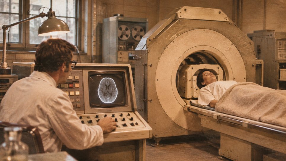
Schulz and colleagues date the first clinical scan to Friday, 1 October 1971, of a living patient's brain, under the supervision of the neuroradiologist James Ambrose. The IEEE milestone plaque ties the image to the same date (as reported by IEEE Spectrum). The patient was a woman with a suspected tumour of the left frontal lobe; according to AuntMinnie Europe she was 41, while Schulz and colleagues say "middle-aged". The results reached Ambrose two days later and showed a cystic lesion in the left frontal lobe. According to Schulz and colleagues the tumour was removed and confirmed as a cystic astrocytoma.
One detail is rough. AuntMinnie Europe gives the same date as the day the scanner was installed and does not state the day of the first patient's scan. So when I say "1 October 1971" I mean the day two sources call the "first image" and one source calls "installation"; I wrote this into the fact-check box.
According to AuntMinnie Europe, the prototype took about four minutes per slice, the slices were about 1 cm thick, and the image was computed from an 80x80 matrix in about 20 minutes on an ICL 1905 mainframe.
An image is also a calculation
On a plain X-ray, different depths of the body overlap in a single shadow. Computed tomography measures how much an X-ray beam is weakened as it passes through the same slice from many angles, and a computer calculates the image of the slice from these measurements. The image is not a photograph taken directly but a solution drawn from measurements. That is why Cormack's mathematics and Hounsfield's machine were two halves of the same idea.
This is a general explanation; I did not read Hounsfield's 1973 system paper for this article.
The announcement: April 1972
According to Schulz and colleagues, the first presentation of clinical data took place on 20 April 1972 at the British Institute of Radiology congress; by then about 70 patients had been scanned. AuntMinnie Europe writes that the first press report appeared in The Times on 21 April 1972. The same review also reports that after the RSNA talk in December 1972 EMI received orders that each required a non-refundable deposit of 100,000 dollars. In 1973 two papers appeared back to back: Hounsfield's description of the system (Br J Radiol 46(552):1016-1022) and Ambrose's clinical application (1023-1047). I know these two citations only from Europe PMC records; the texts were not opened.
The Nobel: half and half
According to the Nobel Foundation's page, the 1979 Nobel Prize in Physiology or Medicine was awarded "for the development of computer assisted tomography" to Allan M. Cormack and Godfrey N. Hounsfield, one half each. Schulz and colleagues give the announcement as 11 October 1979. Oldendorf is not among the laureates. None of the sources I read makes a claim of injustice on his behalf; the bibliography entry says only that the efficiency was not recognised until Hounsfield. I read this not as a claim of injustice but as a record that the prize covered two names. I could not read the Nobel Committee's press release or the text explaining the citation.
The link to cancer
The first patient was scanned for a suspected tumour. According to Schulz and colleagues, Hounsfield's project proposal of October 1968 also spoke of "potential future roles in detecting tumours". The same review writes that body scanners appeared in 1974: Robert Ledley's DISCO system at Georgetown (often cited as the first body scanner), Ohio Nuclear's Delta Scanner prototype at the Cleveland Clinic, and an EMI 5000-series machine at Northwick Park in December 1974. On Day 117 we described the language of staging; I did not separately verify CT's effect on staging and treatment planning for this article, and I do not write it.
Limits and costs
The first limit is the machine itself: only the head was scanned, each slice took minutes, and the calculation needed about twenty minutes. The second limit is access and money. The first orders carried a non-refundable deposit of 100,000 dollars. The model in the Turkey study below takes the cost of a CT device as between 410,000 and 510,000 dollars; this is a model input, not a market price.
The third limit is radiation. CT uses ionising X-rays. In the observational cohort of Pearce and colleagues published in the Lancet in 2012 (178,604 patients, childhood CT scans), the relative risk for leukaemia was 3.18 (95 percent confidence interval 1.46-6.94) for those with a cumulative dose of at least 30 mGy compared with under 5 mGy. The absolute size is much smaller: in children under 10, about one excess leukaemia and one excess brain tumour per 10,000 head CT scans in the 10 years after the first scan. I took these figures from the abstract page; I did not read the full text. Because the study is observational, causal interpretation is limited, and the results concern childhood head CT; they cannot be generalised to adult scans or other body regions.
What remained
- Seeing inside the body slice by slice, without surgery
- The union of measurement and calculation: the image is a solution
- Published mathematics becoming useful through a machine
What fell or weakened
- The assumption that internal structures can be imaged only as overlapping shadows
- The belief that the date an idea is published is the date it becomes useful
- The thought that the cost of every image is only money
From proposals to the scanner and today: 1949-2025. The axis runs from 1945 to 2030. Green marks are dates on which several sources, or a primary citation, agree; blue marks show dates resting on a single source. The amber bar is a period in which sources conflict: Hounsfield joining EMI, between 1949 (Schulz et al.) and 1951 (Nobel biography). Dates known only by year are drawn at mid-year.
CT and a model of national lung cancer screening
I could not verify when the first CT device came to Türkiye; I draw no Turkish link to Hounsfield or Cormack. I report a study that shows how CT reaches the current Turkish debate: Malhan, Göksel, Karadurmuş, Kavaklı, Savaş, Şendur and Şimşek, "The Cost-effectiveness of National Lung Cancer Screening in Türkiye", Turkish Journal of Oncology 2025;40(1), doi:10.5505/tjo.2024.4464. According to the abstract it is a cost-effectiveness model: the NELSON low-dose CT protocol is compared with "no screening" in the 50-74 age range.
The model's outputs: 14 screening rounds; 13,636 stage III-IV cancers found earlier through screening; 7,576 early losses of life prevented; 316,654 additional quality-adjusted life years; 583,840 additional life years gained; 571 dollars per quality-adjusted life year. According to the methods section the model population is 1,019,827 people out of 18,906,477 eligible people aged 50-74. My calculation: 7,576 / 1,019,827 is about 0.74 percent. These are model estimates produced with assumptions, not observed results; I could not read the assumptions from the abstract.
From 1961 to today
What was verified, what stayed uncertain
- Date of the first image: Schulz et al. (J Med Imaging 2021) and the IEEE plaque (via IEEE Spectrum) give 1 October 1971 as the first image; AuntMinnie Europe writes the same date as the installation day and has no scan day for the patient. The first patient's age is 41 (AuntMinnie) or "middle-aged" (Schulz). "Cystic astrocytoma" is only in Schulz et al.; AuntMinnie says "cystic lesion".
- Joining EMI: 1949 (Schulz et al.) versus 1951 (Nobel biography); I could not resolve it.
- Funding: Schulz et al. give 2,500 pounds from the DHSS on 28 October 1968 and a total of 69,000 pounds; the same text calls this "half" of a 10,000-pound request, and the numbers do not match. IEEE Spectrum gives about 40,000 dollars. I could not reconcile them, so I showed the sources rather than state one sum. The story that "Beatles profits financed it" was not verified; IEEE says only that EMI was known for Beatles records and could not fully fund the project.
- Presentation date: 20 April 1972 (Schulz et al.); AuntMinnie gives "April 1972" and The Times report on 21 April.
- Nobel: citation and half shares are from the Nobel Foundation's summary page; the announcement date of 11 October 1979 is from Schulz et al. The press release and the "Perspectives" page could not be opened. Cormack's Nobel lecture page had only the bibliographic details.
- Precursors: the Oldendorf citation is from a bibliography entry; Cormack's volumes and pages were not verified. I did not write the dates for Radon (1917) and Tetel'baum and Korenblyum (1957-58) because I saw them only on Wikipedia.
- 1973 papers and prototype speed: citations are from Europe PMC records, slice and calculation times only from AuntMinnie Europe; I did not write the different figures on Wikipedia.
- Lives of Hounsfield and Cormack: Hounsfield's loss of life on 12 August 2004 is from the Nobel biography; Cormack's years 1924-1998 are from the St John's College page.
- Pearce 2012: from the abstract page (qxmd); volume and pages not verified.
- Malhan 2025: abstract and methods section read (with a summarising tool); the model's assumptions were not read. The arrival of the first CT device in Türkiye could not be verified.
- Yesterday's box: the date of the first clinical image (1 October 1971, installation day in one source) and the sharing of the prize (Cormack and Hounsfield, half each) were verified.
A Break in the Chromosome: Janet Rowley and the Translocation, 1973
Today we looked inside the body slice by slice. Tomorrow we turn to the chromosomes inside the cell: the exchange Janet Rowley saw in leukaemia cells in 1973. I will verify the date of the paper and the role of chromosome banding.
Author's note: What I paid most attention to in this article was not answering "who invented it" with a single name: Oldendorf, Cormack and Hounsfield were different parts, and the Nobel Prize names only two. My second concern was to leave open that one day, such as the date of the first image, may describe different events in different sources (an image or an installation), and to set the benefit of an imaging method beside its radiation cost.
Sources and Further Reading
- Schulz RA, Stein JA, Pelc NJ. How CT happened: the early development of medical computed tomography. J Med Imaging 2021;8(5):052110. doi:10.1117/1.JMI.8.5.052110 (read with a summarising tool; the first 100,000 characters of the text).
- Hounsfield GN. Computerized transverse axial scanning (tomography). 1. Description of system. Br J Radiol 1973;46(552):1016-1022. doi:10.1259/0007-1285-46-552-1016 (Europe PMC record only; the text was not read).
- Ambrose J. Computerized transverse axial scanning (tomography). 2. Clinical application. Br J Radiol 1973;46(552):1023-1047. doi:10.1259/0007-1285-46-552-1023 (Europe PMC record only; the text was not read).
- Nobel Foundation. Summary page of the 1979 Nobel Prize in Physiology or Medicine and biography of Godfrey N. Hounsfield. nobelprize.org (read with a summarising tool).
- Goodrich J. How This Record Company Engineer Invented the CT Scanner. IEEE Spectrum, 12 January 2023 (secondary source; read with a summarising tool).
- AuntMinnie Europe. 50 years of CT scanning approaches (secondary source; read with a summarising tool).
- St John's College, Cambridge. 1979: Allan MacLeod Cormack (1924-1998). joh.cam.ac.uk/node/4401 (read with a summarising tool).
- Oldendorf WH. Isolated flying spot detection of radiodensity discontinuities displaying the internal structural pattern of a complex object. IRE Trans Bio-Med Electron 1961;8:68-72 (via a historyofmedicine.com bibliography entry; read with a summarising tool).
- Pearce MS et al. Radiation exposure from CT scans in childhood and subsequent risk of leukaemia and brain tumours: a retrospective cohort study. Lancet 2012 (via an abstract page; read with a summarising tool; secondary aggregator page).
- Malhan S, Göksel T, Karadurmuş N, et al. The cost-effectiveness of national lung cancer screening in Türkiye. Turk J Oncol 2025;40(1). doi:10.5505/tjo.2024.4464 (read with a summarising tool).
- Mukherjee S. The Emperor of All Maladies: A Biography of Cancer. New York: Scribner; 2010 (general context; the relevant part was not reread for this article).
This article is Day 154 of the "365 Days of Cancer History" series and presents the birth of computed tomography as a historical reading. It is historical-popular content; it is not a diagnosis or a treatment recommendation. Consult your physician for treatment decisions. The sources have been verified; details resting on a single secondary source, on an abstract only, or on a text that could not be read are marked in the fact-check notes.
Was this article helpful?
Your feedback helps us improve our content.