
What Causes a Testicular Lump, and Is It Dangerous? Symptoms, Diagnosis and Treatment Guide

In this article
Testicular Lumps and Scrotal Swelling: Basic Concepts
Scrotal masses refer to any abnormal swelling, lump or hardened tissue within the scrotum, the sac behind the penis. The scrotum contains the testes, responsible for producing sperm and testosterone; the epididymis, where sperm mature; and the network of vessels and nerves supplying the testis (the spermatic cord). Lesions in this region mostly arise directly from testicular tissue, but they may also originate in the epididymis or surrounding membranes. A palpable testicular lump requires serious attention until proven otherwise; however, not every lump means cancer.
- A Painless Lump May Signal Cancer: Testicular cancer usually begins as a painless, hard, board-like lump in the body of the testis. The absence of pain does not mean the condition is harmless.
- Sudden, Severe Pain Is an Emergency: Severe pain in one testis developing within minutes may be caused by the testis twisting around itself (testicular torsion). Surgery within the first 6 hours is essential to avoid losing testicular tissue.
- Most Lumps Are Benign: A substantial proportion of scrotal swellings have benign causes such as fluid accumulation (hydrocele), a spermatic cyst (spermatocele) or dilated veins (varicocele).
- First-Line Diagnosis: Scrotal color Doppler ultrasonography is the gold standard for determining whether a lump is cystic (benign fluid) or solid (suspicious tumor tissue).
Symptoms and Clinical Types of Testicular Lumps
The findings caused by scrotal masses may be acute (sudden) or chronic, depending on the underlying cause:
Physical Changes
A painless, board-hard lump on the surface of the testis, asymmetric enlargement of the entire testis, scrotal fullness and a feeling of heaviness that increases with gravity.
Pain and Radiation
Sudden, sharp testicular pain or a dull, dragging ache radiating toward the groin, lower abdomen and lower back.
Inflammation and Skin Findings
Swelling, redness and warmth of the scrotal skin, with extreme tenderness of the epididymis on touch, usually an infection-related epididymo-orchitis presentation.
Systemic Symptoms
High fever, chills, burning during urination, frequent urination, blood in the urine or, in advanced malignancies, nausea and unintentional weight loss.
Sudden severe testicular pain that is intolerable even to touch, accompanied by nausea and vomiting, requires immediate attendance at the nearest emergency department or consultation with a urologist. This presentation suggests testicular torsion, and minutes are vital to prevent necrosis of the testis.
What Causes a Testicular Lump? Benign and Malignant Causes
Clinical conditions causing masses in the testis and surrounding tissue are examined according to their pathological features:
1. Testicular Cancer
It is generally the most common solid tumor in young men aged 15-35. It is characterized by a hard, usually painless, unilateral mass with an irregular surface arising in the testicular parenchyma. The chance of cure (complete recovery) exceeds 95% when detected early.
2. Spermatocele (Epididymal Cyst)
A well-defined, painless, entirely benign cystic sac just above the testis, formed when clear or milky fluid containing sperm becomes trapped in the epididymal ducts.

3. Epididymitis and Orchitis
Inflammation of the epididymis (the tubular structure storing sperm) or the testis itself. Sexually transmitted microorganisms, such as Chlamydia and gonorrhea, are often involved in young men; urinary bacteria, such as E. coli, or viral agents, such as mumps, are involved in children and older people. It causes severe pain, swelling and fever.
4. Hydrocele
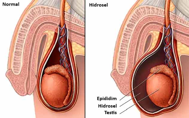
Accumulation of serous fluid beyond physiological limits between the layers of the tunica vaginalis membranes surrounding the testis. This is the most common cause of painless scrotal swelling. It occurs congenitally in babies because the peritoneal canal fails to close, while in adults it develops secondarily after trauma, hernia or infection.
5. Hematocele
Accumulation of blood between the tunica vaginalis layers following trauma, surgery or rupture of a vessel. It causes a hard, tense swelling that is very painful to touch.
6. Varicocele
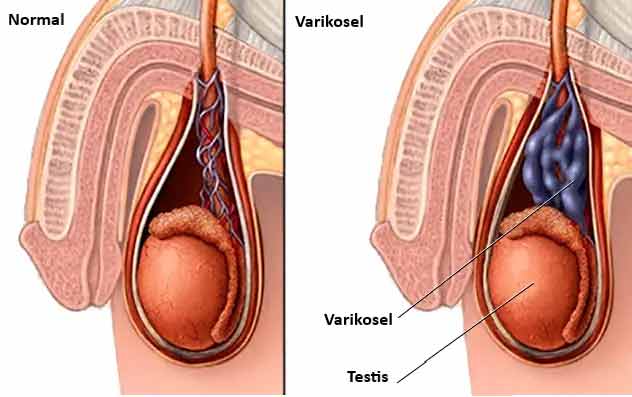
Abnormal dilation and varicosity of the pampiniform plexus veins carrying venous blood from the testis. Because of differences in anatomical venous drainage pressure, it occurs on the left in 85–90% of cases. It feels like a ‘bag of worms’ on examination and may cause chronic pain and male infertility by reducing semen quality.
7. Inguinal Hernia
Protrusion of small bowel loops or omentum into the scrotum because of anatomical weakness in the abdominal wall or a canal remaining open. It becomes more apparent when standing or straining and may recede when lying on the back. It carries a risk of strangulation.

8. Testicular Torsion
Twisting of the spermatic cord around its own axis, obstructing arterial and venous circulation to the testis. It usually develops suddenly during sleep or after exercise in adolescents with a ‘bell-clapper deformity’.
Comparative Differential Diagnosis Table for Testicular Masses
| Mass type | Pain characteristics | Consistency and location | Light transmission (transillumination) | Clinical urgency / approach |
|---|---|---|---|---|
| Testicular cancer | Usually painless (dull heaviness) | Board-hard, solid, within the testis | Does not transmit light (opaque) | Prompt oncological staging and surgery |
| Hydrocele | Painless, feeling of tension | Soft, fluctuant fluid | Transmits light (bright red) | Elective surgery or observation |
| Spermatocele | Painless / mild aching | Epididymal cyst at the upper pole of the testis | May transmit light | Observation if asymptomatic |
| Varicocele | Dull, dragging pain that increases when standing | Veins with a bag-of-worms consistency | Does not transmit light | Semen analysis and microsurgery if needed |
| Epididymitis / orchitis | Acute, severe tenderness | Swollen, edematous, warm and red | Does not transmit light | Medical treatment (antibiotic/anti-inflammatory) |
| Testicular torsion | Sudden, extremely severe pain | A hard testis pulled upward and lying horizontally | Does not transmit light | Emergency surgery (the first 6 hours are the golden period) |
| Inguinal hernia | Fullness-related pain that increases with straining | Soft mass descending from the groin into the scrotum | Does not transmit light (bowel sounds may be heard) | Surgical hernia repair |
Risk Factors for Testicular Masses
The main clinically established risk factors for testicular masses, particularly testicular tumors, are:
- Cryptorchidism (Undescended Testis): Testes that do not descend into the scrotum before birth and remain in the inguinal canal carry, compared with normal testicular tissue, even after surgical descent (orchiopexy), a 4 to 8 times higher risk of testicular cancer.
- Personal and Family Cancer History: A patient who previously had a tumor in one testis has an increased risk of developing a tumor in the other. Individuals with testicular cancer in a first-degree relative, such as a father or brother, have a genetic predisposition.
- Klinefelter Syndrome and Gonadal Dysgenesis: Disorders of embryological testicular development and chromosomal abnormalities increase malignancy risk.
- Childhood Abnormalities: Close urological follow-up during adolescence and adulthood is recommended for children with a history of testicular torsion or an undescended testis.
Diagnostic Methods: Why Is Ultrasound Essential?
The diagnostic process for a man presenting with a testicular lump is conducted carefully, step by step:
The specialist palpates the mass to determine whether it lies within the testicular parenchyma or external structures such as the epididymis. When a bright light is directed at the scrotum in a dark room, fluid masses such as hydroceles transmit the light completely and produce a red glow, while solid tumor tissue and hematoceles block it.
The primary imaging method, without radiation, differentiating masses within minutes with nearly 100% accuracy. It clearly shows whether a mass is solid (tissue suspicious for cancer) or cystic (a benign fluid-filled sac), along with its blood flow.
If ultrasound reveals a solid testicular mass, the following three tumor markers must be measured in blood before surgery: Alpha-fetoprotein (AFP), Beta-hCG and Lactate Dehydrogenase (LDH). These markers play a critical role in monitoring the cancer's type, stage and response to treatment.
When a testicular tumor is confirmed, contrast-enhanced chest, abdominal and pelvic CT is performed to determine whether disease has spread to the retroperitoneal lymph nodes along the posterior abdominal wall or to the lungs.
Treatment Approaches: From Medication to Surgery and Oncology
The treatment protocol depends on the mass's type and pathological origin:
- Infectious Masses: Bacterial epididymitis or orchitis is managed with targeted antibiotics, anti-inflammatory drugs, rest and scrotal elevation (support).
- Benign Masses (Hydrocele, Spermatocele, Varicocele): Small masses that cause no symptoms and do not impair reproductive function may be observed. If there is pain limiting daily life, excessive growth or deterioration in sperm parameters, surgical repair is planned: hydrocelectomy, spermatozolectomy or microscopic varicocelectomy.
- Management of Testicular Cancer:
- Radical Inguinal Orchiectomy: A testis suspected of cancer must never undergo needle biopsy through the scrotum because of the risk of spreading cancer cells to scrotal skin and inguinal lymph nodes. The testis and spermatic cord are removed completely through a small incision in the groin (inguinal approach).
- Chemotherapy and Radiotherapy: Depending on pathology (seminoma or nonseminoma) and stage, platinum-based systemic chemotherapy (the BEP regimen) or radiation oncology comes into play. Testicular cancer has a very high cure rate even with advanced spread.
Testicular Self-Examination (TSE): 3 Minutes Once a Month
Regular self-examination from age 15 is vital for men to become familiar with their genital anatomy and detect testicular cancer early:
- Best Time: During a warm shower or bath. Warm water relaxes the scrotal skin and allows the testes to be felt easily within the sac.
- Inspect in Front of a Mirror: Stand in front of a mirror and look for discoloration, swelling or an obvious deformity of the scrotal skin. It is entirely normal for one testis to hang slightly lower or be very slightly larger than the other.
- Examine Gently with Both Hands: Examine each testis separately. Place your thumb above the testis and your index and middle fingers below it. Gently roll the testis between your fingers.
- Check the Surface and Consistency: Healthy testicular tissue is smooth, oval and slightly firm, with the consistency of a boiled egg. Look for a hard, painless lump or thickening on the body of the testis, even if only pea-sized.
- Distinguish the Epididymis: Feel the soft, slightly ridged cord-like structure running along the back and top of the testis (the epididymis). Do not mistake this normal structure for an abnormal lump.
DROZDOGAN ACADEMY COMMENTARY
The two major mistakes we most often encounter in young men's health are assuming a painless lump is ‘unimportant’ and delaying a medical visit out of embarrassment when testicular pain begins suddenly.
⚡ Why Does Early Diagnosis Save Lives?
Although testicular cancer is the most common solid tumor in young men, it is one of modern medicine's success stories. Even when disease has spread to other organs (metastatic), complete cure rates exceed 90% with appropriate combinations of surgery and chemotherapy. However, reluctance to see a doctor may allow the tumor to spread to lymph nodes and lungs, requiring more intensive treatment.
✅ Remember the Biopsy Rule
While many cancer types are diagnosed by needle biopsy, the firm rule for a testicular mass is: A needle is not inserted through the skin into the testis. Puncturing through the scrotum disrupts the lymphatic barrier and causes cancer to spread to inguinal lymph nodes. Diagnosis is therefore made by approaching through the groin and removing the testis (inguinal orchiectomy).
⚠️ Are Fertility and Hormonal Balance Safe?
Surgical removal of one testis does not by itself eliminate masculinity, erectile function or the ability to have children. The remaining healthy testis alone is more than sufficient for testosterone and sperm production. However, young patients who will receive chemotherapy or radiotherapy after surgery must be offered sperm freezing (cryopreservation) before treatment.
🩺 Our Clinical Approach and Advice
Not every firm area you feel is cancer; it is most likely a cyst or a varicose vein. However, do not try to diagnose a lump yourself. If you notice a change, set embarrassment aside and promptly see a urologist for an ultrasound. Early action protects both your testis and your life.
References and Scientific Guidelines
- European Association of Urology (EAU). Guidelines on Testicular Cancer. EAU Guidelines Office, Arnhem, The Netherlands. Updated 2024.
- National Comprehensive Cancer Network (NCCN). NCCN Clinical Practice Guidelines in Oncology: Testicular Cancer. Version 1.2024.
- American Urological Association (AUA). Diagnosis and Treatment of Early Stage Testicular Cancer: AUA Guideline. J Urol. 2019;202(2):272-281.
- Gordhan CG, Sadeghi-Nejad H. Scrotal pain: Evaluation and management. Korean J Urol. 2015;56(1):3-11.
- Baird DC, Harker GJ, Kabanuk AS. Evaluation of Scrotal Masses. Am Fam Physician. 2019;100(9):535-542.
Sources
Was this article helpful?
Your feedback helps us improve our content.




Reader comments
Share your comment
Your name and comment are published only after administrator approval. Your email address is never shown to visitors.
Be the first to comment.