
Taking Over at 32: Matthew Baillie, the Hunter Legacy and Pathology Made Visible

In this article
Morgagni, whom we discussed yesterday, wrote the birth certificate of pathological anatomy in 1761; but what he wrote was only words. Five volumes, seventy letters, seven hundred cases — all in Latin, all descriptions that readers were expected to visualize in their minds. To see gastric cancer or a pancreatic tumor, one had to read the book and then stand beside an autopsy table, matching the words to the flesh. Thirty-two years later, at an anatomy school on London's Great Windmill Street, a thirty-two-year-old Scottish physician translated the same pathological knowledge into another language : images. His name was Matthew Baillie, and the book he published in 1793, The Morbid Anatomy of Some of the Most Important Parts of the Human Body , was the first pathology textbook organized by organ systemin medical history. Six years later, its visual supplements, published between 1799-1803 — seventy-three high-quality engravings — transformed pathology into a discipline that could be taught, shared and carried across borders. Medical students would now recognize a tumor not by reading , but by seeing .
But Matthew Baillie was more than a pathologist. He was the nephew of perhaps the two most brilliant anatomical geniuses of century 18 — William Hunter and John Hunter . His mother, Dorothea, was the sister of the two Hunter brothers. Baillie's book was therefore the distilled fruit not only of his own years of work, but also of a two-generation Hunter tradition . Thus, 1793 was not merely the publication date of a medical book; it was also the year a legacy changed hands: in that same year, on October 16, 1793, his maternal uncle John Hunter died of a heart attack in London. Thirty-two-year-old Baillie was at his uncle's funeral during the week his book was published. The Hunter family's anatomical legacy now rested on his shoulders.
The Third Generation of an Anatomical Dynasty
Matthew Baillie was born on October 27, 1761 — the year in which Morgagni published De Sedibus— in Shotts, in the Lanarkshire region of Scotland. His father, James Baillie, was a clergyman; his mother, Dorothea Hunter, was the sister of two great names in medicine. His mother's family determined Baillie's destiny.
His older maternal uncle, William Hunter (1718-1783), was one of London's most sought-after obstetricians and a reformer of anatomical education. In 1768, on Great Windmill Street in Soho, he opened a private anatomy school — a model never before seen in medical education. Here, students could dissect cadavers with their own hands, work closely with their teachers and learn from direct examples in anatomical collections. Young physicians came from all over Europe.
His younger maternal uncle, John Hunter (1728-1793), as Önder described on day 65 of the series, was the man who established modern surgery as a science; he also owned a collection of more than 13.000 anatomical and pathological specimens. This collection forms the core of the present-day Royal College of Surgeons of England's Hunterian Museum.
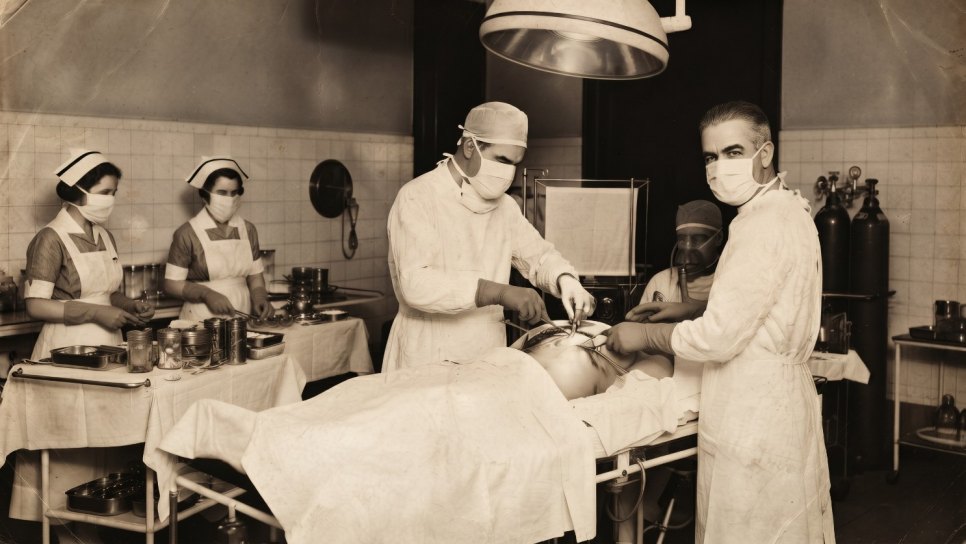
Baillie studied in Edinburgh and at Oxford (Balliol College), then came to London in 1779 and stayed at his uncle's house. William Hunter immediately brought his nephew into the anatomy school; he became a student, an assistant and then a partner. When William died in 1783, Matthew Baillie, aged 22, became the head teacher of the Great Windmill Street Anatomy School that he had inherited. Two years later, at only 24, he was elected to the Royal Society (FRS). He had not yet published his major works, but the European medical world already knew him.
Over the following ten years, Baillie did two things: he taught dissection every week (as Morgagni did in Padua) and systematically recorded every pathological specimen he could obtain. A book was slowly emerging from these records, but it would be different from Morgagni's seventy letters.
1793 — The Morbid Anatomy
Published in 1793, The Morbid Anatomy of Some of the Most Important Parts of the Human Bodyseemed at first glance to be a small book: a single volume, about 350 pages, an unassuming title and no illustrations. But its structure was new.
The Morbid Anatomy — London, 1793
In seventy letters, Morgagni roughly divided the body into anatomical regions — head, chest, abdomen and lower abdomen. Baillie transformed this into a much more systematic organ-based structure : heart, lung, stomach, intestine, liver, spleen, kidney, reproductive organs and brain. For each organ, he first described the diseases to which it was susceptible, then the pathological findings of each disease, followed by example cases. This structure is still the framework of all medical textbooks today. The publication date of 1793 is the birth certificate of the modern organ-based pathology textbook.
The book was a great success. A second edition appeared in 1797 and a third in 1807; it was translated into German, French, Italian and even Russian, and used as a textbook at the medical schools of Edinburgh, Paris and Berlin. But Baillie knew his work was incomplete. Words could describe a tumor's color, consistency and relationship to surrounding tissue only up to a point. The real revolution lay beyond words — in images .
1799-1803 — A Series of Engravings
For six years after the 1793 edition, Baillie worked with the most talented anatomical artists of his time — particularly William Clift, who was John Hunter's assistant and the founding curator of the Hunterian Museum. The aim was to illustrate all the pathologies described in The Morbid Anatomywith high-quality engravings.
Published from 1799 to 1803, this visual supplement appeared as a separate work: A Series of Engravings, Accompanied with Explanations, Which are Intended to Illustrate the Morbid Anatomy of Some of the Most Important Parts of the Human Body. Seventy-three plates. Ten fascicles. Each fascicle contained seven or eight pathological images belonging to a separate organ system. It was the first systematic atlas to show what a lung tumor, a liver metastasis or gastric cancer looked like to the naked eye — transforming knowledge previously confined to individual physicians' anatomical museums into a visual language that could be reproduced in print and carried across borders .
The scale of this revolution may be difficult for a modern reader to grasp. At the end of century 18, a medical student who wanted to learn about a particular tumor had only one option: to see that tumor personally at an autopsy. If no autopsy of that tumor had been performed in the student's city, the student never saw it. However skillful the descriptions in books, it was normal for a surgeon to say, "I have never seen what this looks like in my life." Baillie's atlas ended this situation. A student in Edinburgh, one in Padua and one in Boston — all looked at the same seventy-three plates, learning the same tumors through the same images. Pathology ceased to be a national science and became an international language.
Lung, Stomach, Brain, Liver — Visually Documented Tumors
The cancers to which Baillie gave particular attention in both his 1793 text and his 1799-1803 atlas were as follows:
- Lung cancer: In his atlas, Baillie was the physician who documented a solid, fungating or scirrhous mass in the lung with a systematic visual signature for the first time . His cases included masses growing around the bronchial tree, tumor infiltration involving the pleura and enlargement of mediastinal lymph nodes. The relationship between smoking and lung cancer had not yet been formulated — John Hill sensed that connection in 1761 (Day 63), and Doll-Hill proved it in 1950 (Day 129); but Baillie was the first physician to draw the anatomical picture of lung cancer in the body.
- Gastric cancer (scirrhous pylorus): A description of a thick, fibrous tumor in the pyloric region, narrowing the lumen. The relevant plate in his atlas is the first record to visually confirm the relationship between a patient's vomiting and weight-loss symptoms and this anatomical finding. Baillie showed through engraving what Morgagni had described in writing.
- Pituitary tumors: A description of soft, compressive masses at the base of the brain, in the sella turcica region. The term "pituitary adenoma" did not exist at the time; Baillie called these lesions "tubercles of the brain base". His description, however, corresponds exactly to the classic anatomical appearance of a modern pituitary macroadenoma. It was a conceptual preparation for the endocrinological picture that Cushing (to be covered on Day 117) would solve a century later.
- Liver tumors — distinguishing primary from metastatic disease: In his atlas, Baillie proved the distinction initiated by Morgagni visually . On the same plate, the single-focus tumor image of primary liver cancer compressing surrounding tissue was printed beside the multifocal, scattered nodular image of metastatic liver involvement. This is the visual ancestor of the distinction we still use in modern radiology reports.
- Testicular and uterine tumors: The first systematic visual records of cancers of the reproductive organs are also in Baillie's atlas.
These engravings in Baillie's atlas became the standard referencefor European medical education for the next century. If a student in Berlin had not seen gastric cancer in 1850, they consulted the plate in Baillie's atlas. When a physician in Edinburgh examined a patient with suspected pituitary tumor in 1880, the atlas's description was still vivid in their mind.
The Establishment of Anatomical Museums as a Scientific Enterprise
Baillie's second major contribution was a museum, not a book. When his maternal uncle John Hunter died in 1793, he left an enormous collection of 13.687 anatomical and pathological specimens. This was a scientific data archive on a scale never before seen in English history — cancer specimens, malformations, normal anatomical variants and experimental surgical specimens. Despite his wife's warnings, Hunter did not want the collection to be left on its own, but his will was complicated.
Baillie and William Clift organized the collection's sale to the British government . In 1799, the government purchased the collection for 15.000 pounds sterling (approximately 2 million pounds sterling at today's value) and transferred it to the Royal College of Surgeons in 1800. The collection moved first to Lincoln's Inn Fields, then to the Royal College's present building. Today's Hunterian Museum is the ancestor of modern medical collecting — and the principal figure responsible for this institution's survival was Matthew Baillie.
This has great significance for the history of cancer. A museum is not merely an aesthetic or historical place; it is an archive of scientific data. A pathologist in the 1850s did not have to settle for the description in Baillie's atlas — they could go to the Royal College and examine the original Hunter specimen. This is the conceptual origin of modern biobanks and tumor tissue archives . Today, Memorial Sloan Kettering's 50-year tumor archive, M.D. Anderson's partnership with Foundation Medicine and the biobank of Hacettepe Oncology Institute in Turkey are all extensions of the tradition that Baillie and Clift began in 1799.
👑 AN ASIDE: PHYSICIAN TO ROYALTY AND A POET
From 1799 onward, Baillie became London's most sought-after physician in internal medicine. In 1810, he was appointed chief physician to King George III — leading the team that attended the king during his "madness" (interpreted in modern debate as porphyria or bipolar disorder). He later also served as physician to Lord Byron and Caroline, Princess of Wales . His handwritten examination notes appear in the final moments of many members of the royal family. He earned a considerable fortune from his practice during his lifetime and donated a substantial part of it to the Royal College of Physicians, its library and scholarship funds. In 1817, he retired from active practice and withdrew to the countryside. He died in Gloucestershire on September 23, 1823 — leaving both a pathology textbook that had gone through three editions and the founding story of one of British medicine's most important museums.
The 230-Year Echoes of an Atlas
📖 THE VISUAL LANGUAGE OF PATHOLOGY — FROM BAILLIE TO TODAY
The traces of that book printed in 1793 extend to today's digital pathology platforms. When a pathologist at Memorial Göztepe discusses a biopsy decision with a colleague in Stockholm, they are actually the latest link in the principle that Baillie established 230 years ago: "Pathology is a visual language." The original specimens still displayed at the Hunterian Museum are the ancestors of modern biobanks. The Royal College of Surgeons' collection from century 18 is the first example of an infrastructure now reproduced at every center conducting oncology research worldwide.
🔍 A BRIEF FACT-CHECK NOTE
- Birth and death: October 27, 1761, Shotts (Scotland, Lanarkshire) — September 23, 1823, Duntisbourne House (England, Gloucestershire), aged 61
- Mother: Dorothea Hunter — sister of William Hunter (1718-1783) and John Hunter (1728-1793)
- Education: University of Edinburgh, followed by Balliol College, Oxford
- Great Windmill Street Anatomy School: Took over in 1783, aged 22; led it until 1799
- Royal Society (FRS) fellowship: 1790
- The Morbid Anatomy: London, 1793; three editions (1793, 1797, 1807); translated into many languages
- A Series of Engravings: London, 1799-1803, ten fascicles, seventy-three plates; collaboration with the artist William Clift
- Hunter collection: 13.687 specimens; sold to the government in 1799 for 15.000 pounds sterling; at the Royal College of Surgeons from 1800 onward
- Royal physician: Chief physician to King George III from 1810; also physician to Lord Byron and Caroline, Princess of Wales
Marie Antoinette's Mother: Empress Maria Theresa and Illness at the Habsburg Court
From the laboratory of pathological anatomy to the royal bedchamber... Maria Theresa, one of the most powerful women in Europe in century 18, is among the first cases in which a long-discussed posthumous "suspicion of cancer" can be traced in court documents. Marie Antoinette's mother, a woman who shaped political decisions during the Enlightenment: what uncertain illness accompanied her final years? Tomorrow we open a thematic dossier on court physicians' records, the medical limitations of the period and the concept of "royal illness."
Author's note: The Baillie quotation in this article is adapted from the A Series of Engravingspreface of 1799 into Turkish; it is a conceptual summary, not a literal translation of the original English text. Baillie's descriptions of cancer do not correspond one-to-one to today's concept of histopathological subtypes — the morphological language of century 18 (scirrhous, fungous, melisseros, encephaloid) was a classification based on consistency and color. Its transformation into modern molecular and histological classification will be discussed in the coming days of the series in the articles on Müller (Day 81), Virchow (Day 87) and Waldeyer (Day 90).
Sources and Further Reading
- Baillie M. The Morbid Anatomy of Some of the Most Important Parts of the Human Body. London: J. Johnson and G. Nicol, 1793. (First edition.)
- Baillie M. A Series of Engravings, Accompanied with Explanations, Which are Intended to Illustrate the Morbid Anatomy of Some of the Most Important Parts of the Human Body. Illustrators: William Clift and James Heath. London: W. Bulmer & Co., 1799-1803. (Ten fascicles; seventy-three plates.)
- Wardrop J. Memoirs of the Life and Writings of Matthew Baillie, M.D. London: J. Murray, 1825. (A biographical source published two years after Baillie's death.)
- Crainz F. The Editions and Translations of Dr. Baillie's "Morbid Anatomy". Medical History 1982;26(4):443-452.
- Russell KF. Matthew Baillie F.R.S. (1761-1823). Medical History 1972;16(1):1-22.
- Long ER. A History of Pathology. New York: Dover Publications, 1965. (The place of the Morgagni-Baillie transition in the history of pathology.)
- Moore W. The Knife Man: The Extraordinary Life and Times of John Hunter, Father of Modern Surgery. New York: Broadway Books, 2005. (The history of the Hunter family and the intellectual environment in which Baillie grew up.)
- Cooke AM. A Forgotten Sketch of Matthew Baillie. Journal of the Royal College of Physicians of London 1979;13(3):189-191.
- Royal College of Surgeons of England. The Hunterian Museum: A Guide. London: Royal College of Surgeons, various editions. (The process by which Baillie transferred John Hunter's collection to the institution.)
- Hajdu SI. A note from history: landmarks in history of cancer, part 2. Cancer 2011;117(11):2811-2820. doi:10.1002/cncr.25855
This article is day 69 of the History of Cancer in 365 Days Series. The subsequent links in the chain from the visualization of pathological anatomy to modern digital pathology — Bichat (Day 73), Müller (Day 81), Virchow (Day 87), Waldeyer (Day 90) and modern AI-assisted pathology (Day 326) — will be covered in the coming days of the series. The article is written from a popular historical perspective and provides no guidance on medical treatment.
Was this article helpful?
Your feedback helps us improve our content.


Reader comments
Share your comment
Your name and comment are published only after administrator approval. Your email address is never shown to visitors.
Be the first to comment.