
What Are Calcifications in the Breast and on Mammograms? Should You Be Concerned?

In this article
Breast calcifications are small calcium deposits in breast tissue. They are usually seen on mammograms and occur most frequently in women over age 50. Breast calcifications are typically noncancerous, or benign. However, certain forms can sometimes indicate breast cancer.
In this article, we discuss the causes of breast calcifications and what may happen if they appear on a screening test.
WHAT Are Breast Calcifications?
Calcifications are small calcium deposits that develop in the breasts. They do not cause signs or symptoms, and a doctor cannot diagnose them during a routine breast examination.
Instead, they usually appear as white spots on a mammogram , an X-ray image of the breast. Other breast scans, such as ultrasound or magnetic resonance (MR) scans, do not show calcifications.
Calcifications are more likely to develop after menopause.
Most breast calcifications are benign
However, clusters of calcium deposits can also be an early sign of breast cancer. Some research reports that calcification in the breast is the only warning sign of cancer in 12.7 to 41.2% of women in screening programs.
Although calcifications do not turn into cancer , they may suggest that an underlying process is occurring that could be associated with breast cancer.
What Are the TYPES of Breast Calcifications?
Two types of breast calcifications can be seen on mammograms: macrocalcifications and microcalcifications . Some women may have both types.
Macrocalcifications
Macrocalcifications appear as large white spots occurring randomly in breast tissue. They are greater than 0,5 millimeters (mm) in diameter.
Macrocalcifications are very common, and radiologists consider these structures benign. As a result, people with macrocalcifications in the breast generally do not need close follow-up testing.
Microcalcification
Microcalcifications appear as small white specks or grains. Many cases of microcalcification are benign. However, if they have certain characteristics, they may indicate breast cancer.
Microcalcifications require further investigation if they have the following characteristics:
- Less than 0,5 mm in diameter
- A tendency to cluster in one area of the breast
- Variations in size and shape
- A cluster usually consists of five or more calcifications, but this number is not fixed. Loose clusters are more likely to be benign than compact/tight clusters.
Calcifications Outside the Main Breast Tissue
Sometimes a mammogram may show calcifications outside the main breast tissue. These may appear in the skin or inside blood vessels.
Deodorant or powder residue on your skin may cause white spots to appear on X-ray results.
If the radiologist confirms that the calcifications are located outside the breast tissue, these types of calcification do not require further testing.
WHAT CAUSES Calcifications / Calcium Deposits in the Breast?
Calcification in breast tissue may develop for the following reasons:
- Trauma from an injury or surgery. Calcifications are common after breast surgery.
- Infection
- Formations similar to atherosclerosis in blood vessels
- Cysts, which are fluid-filled sacs in the breast
- Mammary duct ectasia or blocked milk ducts
- Fat necrosis or dead fat cells
- Fibroadenoma, a benign breast tumor
- For breast cancer, radiation therapy
- Ductal carcinoma in situ (cancerous changes in the cells lining the milk ducts, very early-stage breast cancer)
- Invasive breast cancer (classic breast cancer)
- You may also be interested in: 6 Risk Factors for the Progression of Ductal Carcinoma In Situ (DCIS) to Breast Cancer
Any of the factors above can cause breast calcifications to develop.
Other factors that increase the risk include:
- A personal history of breast cancer
- A family history of breast cancer
- A hereditary predisposition to breast cancer, such as having BRCA1 and BRCA2 mutations
Note: Deodorant, lotions or powder may appear as calcifications on a mammogram. People should therefore avoid applying any skin products before screening.
Eating calcium-rich foods does not cause breast calcifications!
How Are Breast Calcifications DIAGNOSED?
When Is Further Investigation Necessary?
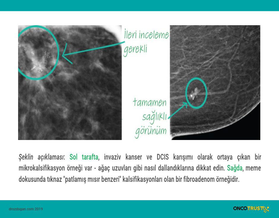
If calcifications appear on a mammogram, the radiologist decides whether further investigation is needed. In general, they do not consider macrocalcifications suspicious.
However, if large clustered calcifications or microcalcifications are detected, the radiologist may recommend further tests.
If the calcifications are considered suspicious for cancer, the following may be done:
- Another mammogram to view the calcifications more closely,
- Comparison with earlier mammogram images to see whether the characteristics of the calcifications have changed,
- Recommending screening every 6 months to check for changes in the calcifications,
- Recommending a breast MR scan,
- Breast biopsy may be performed,
TREATMENT of Breast Calcifications
A doctor can discuss possible treatments for medical conditions causing breast calcifications. Benign calcifications do not require treatment. However, if calcifications are associated with another medical condition — mammary duct ectasia, for example — the doctor will provide the person with more information about the condition and available treatments.
If calcifications indicate breast cancer, the person will of course need treatment to stop the cancer from spreading and kill cancer cells. The type of treatment a person needs depends on the type, size and stage of the breast cancer and other factors.
*
Sources
The importance of early detection of calcifications associated with breast cancer in screening.
Breast Cancer Res Treat. 2018
2. Yojana V Nalawade.
Evaluation of breast calcifications.
Indian J Radiol Imaging. 2009 Nov.
Was this article helpful?
Your feedback helps us improve our content.


Reader comments
Share your comment
Your name and comment are published only after administrator approval. Your email address is never shown to visitors.
Be the first to comment.