
Astley Cooper: The Anatomical Birth of Breast Surgery and the Unwritten Cancer Book

In this article
“Cooper’s Ligaments” and the Anatomical Birth Certificate of Modern Breast Surgery — But Also the Story of an Unpublished Second Volume
In February 1829, a quarto volume appeared in London, published by Longman, Rees, Orme, Brown and Green: Illustrations of the Diseases of the Breast — Part I. Its author was the most famous surgeon of his era, the legendary anatomist of Guy’s Hospital, granted a baronetcy by King George IV: Sir Astley Paston Cooper. 89 pages of text, nine hand-colored plates, and the distilled essence of decades of dissection experience. Yet the most surprising part of this story lies in an often overlooked fact: Part I, published in 1829, covered only benign breast diseases; the Part II Cooper promised — a systematic examination of breast cancers — was never published. When Cooper died in 1841, he was the richest and most famous surgeon in the world; he was buried as a king’s doctor, but the drafts of this second volume were left on his desk. In this article, we will examine Cooper’s life, his lasting contributions to breast anatomy (the ligaments that still bear his name and the fibrocystic condition known as Cooper’s disease), what the book of 1829 actually did and did not do, where the era’s scirrhus/encephaloid classification came from and where it went, and Cooper’s true place in the history of oncology. Let us state this at the outset: Astley Cooper is notthe father of the modern classification of breast cancer. He is, however, the man who established the anatomical foundations of modern breast surgery, and this distinction deserves careful preservation when writing the history of cancer.
23 August 1768: The Fourth Son of a Norfolk Pastor
Astley Paston Cooper was born on 23 August 1768 at Brooke Hall, in the village of Brooke near Norwich. His father, Dr. Samuel Cooper, was an Anglican clergyman: rector of Morley and Yelverton and vicar of Great Yarmouth. His mother, Maria Susanna Bransby, was a writer of children’s stories and epistolary novels who achieved considerable success in her day — her books are said to have sold more than her husband’s sermons. The family, his childhood household, also maintained a tradition of descent from Sir Isaac Newton ; although far from certain, this claim illustrates how the educated classes of Norfolk at the time constructed their identity.
Astley was the family’s sixth child and fourth son. His father’s remark became legendary: “He is a mischievous boy, but despite his mischief, I have no doubt that he will be a character who shines in the future.” In early childhood, he was not sent to school like an ordinary rural boy, but was educated by his family and the village teacher. When he was around ten, he witnessed a friend’s death after the friend’s popliteal fossa was crushed beneath a cart. Cooper later said this event determined the direction of his life: “That day I became convinced of how valuable a member of society a good surgeon is, and how great a curse an ignorant surgeon is."
At 16 he was sent to London. His mother’s brother, William Cooper, was a senior surgeon at Guy’s Hospital; the family connection opened the way. Astley first began at Guy’s with his maternal uncle in August 1784, then entered the apprenticeship and household of St Thomas’ Hospital surgeon Henry Cline(1750-1827). Cline was a crucial mentor: an accomplished anatomist and surgeon, and also an intellectual close to the liberal-radical political movements of the time. As a young man, Cooper also immersed himself in this radical political atmosphere, during the years when the French Revolution of 1789 resonated widely in London. In later years, however, he withdrew entirely from politics, preferring the social security offered by his profession to political reformism.
The main intellectual influence came from the lectures of the greatest anatomist and surgeon in London at the time, John Hunter(1728-1793). Cooper attended Hunter’s lectures again, year after year, because they were not repetitions: every year they were updated with Hunter’s new observations and experiments. Once, when Cooper reminded Hunter that “last year you said precisely the opposite on this subject”, Hunter’s reply was itself a lesson: “I knew less then.” This sentence summarizes the epistemology of modern science.
Anatomy Demonstrator at 21, Guy’s Hospital Surgeon at 32
Cooper’s career rose extraordinarily quickly:
CHRONOLOGY OF ACADEMIC ADVANCEMENT
- 1787-1788: A year at Edinburgh Medical School. Edinburgh was the strongest anatomical-surgical school in the British world at the time; figures such as William Cullen and Joseph Black were still alive.
- 1789: At 21, Anatomy Demonstrator at St Thomas’ Hospital — the person conducting dissection classes.
- 1791: Joint lecturer in anatomy and surgery with Cline.
- 1791: In the same year, he married the daughter of London merchant Thomas Cock, Ann Cock . Their only child died at a young age, the great tragedy of Cooper’s private life.
- 1793-1796: Anatomy lecturer at the Royal College of Surgeons; one of his duties was the public dissection of criminals executed at the Old Bailey.
- 1800: At 32, he succeeded his maternal uncle William Cooper as surgeon at Guy’s Hospital . In the same year, he was elected a Fellow of the Royal Society.
- 1801-1802: The Royal Society’s Copley Medal — for two papers on the effects of perforations of the eardrum (tympanic membrane). This was a lasting contribution by Cooper to otology.
- 1817: For the first time in history, he performed ligation of the abdominal aorta for an aneurysm. The patient died a few days later, but the procedure itself was a technical milestone.
- 1820: He removed an infected sebaceous cyst from the scalp of King George IV.
- 27 July 1821: In return for this procedure, he was granted the title of Baronet of Gadebridge . Cooper was now Sir Astley Cooper.
- 1825: Two years before his wife Ann died, he retired from his active position at Guy’s because of health problems, although he continued as a consulting surgeon.
- 1827 and 1836: President of the Royal College of Surgeons — two separate terms.
- 1828: George IV’s Sergeant-Surgeon— later he would also serve as Sergeant-Surgeon to William IV and the young Victoria. Thus, chief surgeon to three successive British monarchs.
- 12 February 1841: He died at his home on Conduit Street, aged 72.
Economic success accompanied Cooper’s professional success. Between 1815-1820, his annual income ranged from 21.000 to 22.000 pounds — the equivalent of millions of pounds today. Following recovery from an illness, he was rewarded with a baronetcy by the king; he had also risen from his beginnings as a village boy to a mansion with his own workshop-laboratory in London’s most expensive district.
An important note: Cooper’s passion for anatomy also brought with it the era’s shortage of bodies for dissection. Before the Anatomy Act of 1832, legal channels for obtaining cadavers in England were extremely limited; in practice, surgeons had to work with “resurrection men” — grave robbers . Cooper’s connection with this network is documented; he is known to have been an active buyer himself and even to have made annual payments to particular grave robbers. On the one hand, this was common practice in his era; on the other, it is disturbing by our modern ethical standards. Cooper actively advised Parliament on the passage of the Anatomy Act of 1832, thus also contributing to legalization of the system.
Only Benign Diseases — A Promised but Unwritten Second Volume
Published in London in February 1829, the book is one of the major works of Cooper’s mature years. The edition from Longman and his four partners contained 89 pages of text and 9 hand-colored plates. Each plate had an accompanying explanatory leaf. The volume was quarto, a large format, and its production quality was outstanding for the era.
But the most crucial point here is the fact often overlooked or misrepresented: This book was planned in two volumes, and Part I was devoted only to benign breast diseases. According to Cooper’s own preface, the first volume dealt with diseases that “do not arise from a disordered state of the system, produce no dangerous structural effects, do not contaminate neighboring parts and do not affect distant structures”. The medical-philosophical distinction underlying this sentence is important: within the Hippocratic-Galenic framework of his era, Cooper accepted the distinction between “diathesis” (a systemic disorder of the body) and “local disease”. Benign diseases were local; malignant diseases were systemic. He therefore conceived two systematically separated volumes .
The ten chapters of Part I covered breast anatomy, development, lactation, mastitis, abscesses, fibrocystic changes (what Cooper called “hydatid disease”), simple cysts, fibroadenomas, neuralgia (breast pain) and other benign conditions. Most plates showed specimens from Cooper’s personal dissection museum.
📖 PART II — THE UNWRITTEN SECOND VOLUME
The “second volume on malignant diseases” promised in Cooper’s preface was never published. In the 1830s, Cooper worked on other projects: Diseases of the Testis (1830), The Anatomy of the Thymus Gland (1832), and finally his return to breast anatomy in 1840, On the Anatomy of the Breast (1840). This final work entered history as the definitive classic text on breast anatomy; the structures still known as “Cooper’s ligaments” were described in detail in this book. But none of these was the planned Part II on “breast cancers”.
Why did Cooper not complete this second volume? Historical sources offer no definitive answer. Possible explanations: (1) scientific understanding of malignant breast disease was still unclear at the time, and Cooper did not wish to publish inadequate data as a matter of epistemic honesty; (2) aging and health problems — Cooper experienced increasingly severe chronic ailments in his final 15 years; (3) shifting priorities — he considered completing breast anatomy itself more fundamental than classifying disease. Whichever is correct, the result is this: Cooper’s planned contribution to breast cancer classification in the history of oncology remained incomplete.
The Misattribution of a Classification: The General Framework of Morphological Pathology Early in Century 19
Popular sources on modern cancer history sometimes state that “Astley Cooper divided breast cancer into scirrhus and encephaloid”. This statement is wrong, or at least extremely oversimplified. Scirrhus (hard cancer) and encephaloid (soft cancer, “brain-like”) are not terms unique to Cooper. They are morphological descriptions widely used throughout European surgical-pathological literature from late in century 18 to the middle of century 19.
The terms have very ancient origins. Scirrhus comes from the Greek σκίρρος, meaning “hard stone”; in classical Galenic medicine, Galen and Ibn Sina used it to describe a form of tumor. Encephaloid(brain-like) developed as a systematically used term in the French school of pathology late in century 18, particularly with Marie François Xavier Bichat (Day 73) and the Paris school that followed him. Bichat’s work of 1800, Anatomie générale, introduced this distinction into medical language by separating “medulla-like” tumors (soft, gray and watery in appearance) from hard “squirrus”.
Throughout the early years of century 19, the morphological scirrhus/encephaloid distinction stood in place of modern subtype classification. Before microscopic examination, however, it was a distinction made only with the naked eye and by palpation . Hard and slow-growing = scirrhus; soft, brain-like and fast-growing = encephaloid. The Britannica Encyclopaedia of 1911 still records this distinction: “Scirrhus is notable for its hardness, which is due to the large quantity of fibrous elements and the relatively small proportion of cellular elements. It grows slowly but tends to spread and ulcerate. Its most common site is the female breast."
Modernization of this classification had to wait for the microscope to enter clinical pathology practice. Johannes Müllerreconsidered the scirrhus/encephaloid distinction at the cellular level in his 1838 work, Über den feineren Bau und die Formen der krankhaften Geschwülste(On the Fine Structure and Forms of Malignant Tumors), the foundational book of microscopic cancer pathology. Rudolf Virchowtransformed the classical morphological categories in his work of the 1850s within the framework that “cancer is a disease of the cell”. Carcinoma (an epithelial tumor) and sarcoma (a connective tissue tumor) came to be distinguished during this microscopic era, rather than through Astley Cooper’s work.
THE DISTINCTION WIDELY USED IN 1829
Scirrhus: Hard, fibrous, slow-growing cancer (its modern counterpart is mostly invasive ductal carcinoma). Encephaloid (medullary): Soft, fast-growing cancer with the appearance of brain tissue (its modern counterpart is medullary carcinoma + certain high-grade forms). The distinction rests on the naked eye + touch.
THE MODERN DISTINCTION IN 2026
Luminal A, Luminal B, HER2-enriched, triple-negative (TNBC) — molecular subtypes. ER, PR and HER2 immunohistochemistry + Ki-67 index + a recurrence score based on 21 genes (Oncotype DX) + genomic testing (PAM50). 200 years beyond Cooper’s era, yet an extension of the same basic logic: “Not all breast cancers are the same; subtypes behave differently.”
The important point is this: scirrhus and encephaloid categories were not discussed in Cooper’s Part I, written in 1829, because that volume was devoted to benign diseases. Cooper used these terms in other works and clinical reports, but he did not create them and did not publish a systematic monograph on them. Calling Cooper “the father of breast cancer classification” in the history of oncology — an error sometimes found in popular sources — is historically indefensible.
1840: On the Anatomy of the Breast — The Definitive Classic Text
Cooper’s truly monumental contribution to breast history was the work he published eleven years after 1829, only a year before his death, in 1840: On the Anatomy of the Breast. This is the definitive classic text on the glandular anatomy of the breast, a foundational reference still cited by medical textbooks discussing breast anatomy today.
WHAT COOPER FOUND INSIDE THE BREAST
Cooper systematically described the lobular-lobar-ductal-acinar organization of the breast gland, the average number of lobes per lobule, the distribution pattern of the major lactiferous ducts at the nipple, and the fibrous connective tissue structures attaching glandular tissue to the skin.
These fibrous connective tissue structures are still called “Cooper’s ligaments” (Cooper’s suspensory ligaments) today. They are thin fibrous bands connecting the dermis to the pectoral fascia; they provide the breast’s shape and firmness. They stretch with age and gravity, providing the anatomical explanation for the mechanism of ptosis, or sagging, familiar in aesthetic plastic surgery.
The principal importance of Cooper’s ligaments in clinical oncology lies elsewhere: Breast cancer causing fibrotic retraction of Cooper’s ligaments leads to visible skin dimpling or an “orange peel” (peau d’orange) appearance. This physical examination finding — the first step in the modern clinical breast examination — is the direct clinical legacy of Cooper’s anatomical work. When a surgeon or physician sees this retraction of the skin, what they know without seeing is that an underlying tumor is pulling on the connective tissue structure Cooper described in 1840.
Cooper’s other enduring breast eponyms:
👨⚕️ COOPER EPONYMS IN ANATOMY AND PATHOLOGY
- Cooper’s ligaments (Cooper's suspensory ligaments): The fibrous connective tissue structures described above. A fundamental anatomical reference for modern breast surgery.
- Cooper’s disease (Cooper's disease): Benign cystic disease of the breast — today’s fibrocystic breast disease. Cooper described it as “hydatid disease”, meaning “cystic”.
- Cooper’s neuralgia (Cooper's neuralgia of the breast): Idiopathic chronic pain of the breast, corresponding to modern mastalgia.
- Cooper’s testis (Cooper's testis): A male genitourinary anatomical structure, an eponym associated with the testis.
- Cooper’s fascia (Cooper's fascia): The fascial layer around the spermatic cord in the inguinal region.
- Cooper’s hernia (Cooper's hernia): A type of perineal/crural hernia variant.
- Cooper’s pubic ligament (Cooper's pubic / pectineal ligament): A strong fibrous band on the upper pubic ramp, still important in hernia surgery today, especially in femoral hernia repair.
- Cooper’s strips: Tendinous fibrous bands, an anatomical variant.
Thus, Cooper’s true role in the history of breast oncology is as an anatomist, rather than a classifier. Everything modern breast surgery will do — from Halstead’s radical mastectomy in 1894 to Veronesi’s lumpectomy in 1969, Krag’s sentinel lymph node biopsy in 1993 and today’s oncoplastic surgery — takes place on the breast anatomy Cooper mapped. When a surgeon touches the breast, knowingly or unknowingly, they follow the path of Cooper’s atlas of 1840.
The Context of Mastectomy Before Anesthesia
Breast cancer practice in Cooper’s time was unimaginably different from today. A few fundamental facts:
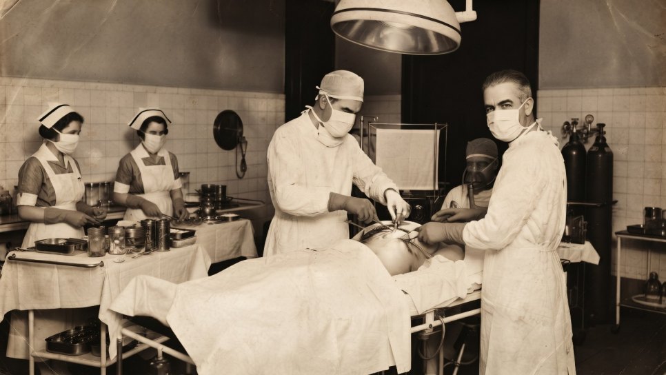
First — there was no anesthesia. Cooper died in 1841; the first public demonstration of ether anesthesia was performed 5 years later, on 16 October 1846, by William T.G. Morton at Massachusetts General Hospital in Boston (Day 89 of the cancer history series). Thus, in Cooper’s era, every breast operation was performed on a fully conscious patient . The criteria for surgical success were different: speed, decisiveness, bleeding control and the patient’s capacity to bear pain. The famous letter describing the mastectomy of Cooper’s contemporary Frances Burney on 30 September 1811 (Day 71) is the earliest first-person source for this pain.
Second — there was no antisepsis. Joseph Lister’s carbolic acid antisepsis would arrive in 1867. In Cooper’s era, surgical wound infections (sepsis, “hospital gangrene”, erysipelas) were extremely common; mortality after mastectomy was around 40-50%, often due to infection rather than surgical adverse effects.
Third — there was no systemic treatment. The assumption that breast cancer was a local disease dominated until 1894, with Halsted’s radical mastectomy paradigm. Systemic chemotherapy came after the 1940s, hormonal therapy after the 1970s, and targeted therapy after 1998 (trastuzumab). In Cooper’s era, the idea that “breast cancer is a systemic disease” either did not exist or was a mystical Hippocratic concept within the framework of diathesis, a systemic disorder.
Together, these three deficiencies meant that treatment options for breast cancer in Cooper’s era were extremely limited: surgical resection in early stages, with a high risk of operative death, and palliative approaches in advanced stages (opium, herbal laxatives, nutritional support and religious-spiritual support). Survival was extremely poor.
A clinical observation: The fact that Part I of Cooper’s book of 1829 was devoted to benign diseases was not only an epistemic choice but also a practical necessity. The results of surgical/local treatment for benign diseases were clear; for breast cancer, they were uncertain and mostly disastrous. As a surgeon, Cooper preferred to write about benign conditions because he had meaningful things to say in that area. The fact that the planned Part II on malignant diseases remained unwritten may partly reflect Cooper’s scientific integrity: he preferred not to leave a classic text in a field where he had nothing definite to say.
FROM COOPER TO TODAY — THE BREAST ONCOLOGY CHAIN
INFORMATION VERIFIED IN THIS ARTICLE
- Birth and death: 23 August 1768, Brooke Hall, Norfolk — 12 February 1841, Conduit Street, London. Died aged 72.
- Father: Dr. Samuel Cooper (Anglican clergyman, rector of Morley-Yelverton)
- Mother: Maria Susanna Bransby (writer of children’s stories and epistolary novels)
- Maternal uncle: William Cooper (Guy’s Hospital surgeon, family connection)
- Mentors: Henry Cline (St Thomas’) + John Hunter (lectures)
- Education: 1784 Guy’s Hospital → 1787-1788 Edinburgh → St Thomas’ Hospital
- 1789: Anatomy Demonstrator at St Thomas’ at 21
- 1791: Joint lecturer in anatomy and surgery with Cline; marriage to Ann Cock in the same year
- 1800: Guy’s Hospital surgeon + Fellow of the Royal Society
- 1801-1802: Copley Medal (for otology studies)
- 1817: The first ligation of the abdominal aorta in history
- 1820: Removal of a sebaceous cyst from King George IV
- 27 July 1821: Baronet of Gadebridge, Hertfordshire (London Gazette Issue 17720)
- 1825: Retirement from the active Guy’s position, remaining as consulting surgeon
- 1827 and 1836: President of the Royal College of Surgeons (two terms)
- 1828: Sergeant-Surgeon to George IV
- February 1829: Illustrations of the Diseases of the Breast, Part I published (Longman, Rees, Orme, Brown and Green; 89 pages + 9 color plates)
- Important correction: Part I was devoted only to benign diseases; Part II (malignant diseases) was never published
- 1840: On the Anatomy of the Breast published — the definitive work describing Cooper’s ligaments
- Famous student: Poet John Keats (Guy’s Medical School, licensed in 1816; notes from Cooper’s lectures have been preserved)
- Scirrhus/encephaloid classification: Not unique to Cooper; morphological terms widely used by Bichat (1800) and in the era’s European surgical-pathological literature. Microscopic cancer pathology arrived with Müller (1838) and Virchow (1850s).
Robert Liston and the “Half-Minute Mastectomy” — The Aesthetics of Surgical Speed Before Anesthesia
We move from Cooper’s London to Edinburgh, from Cooper’s baronetcy to the generation that followed. Robert Liston (1794-1847), a Scottish surgeon and the fastest operator of his era, is said to have completed a mastectomy in 30 seconds. In the age before anesthesia, speed was kindness: the most powerful means of shortening the time a patient endured pain. On 21 December 1846, Liston would perform Britain’s first operation under ether anesthesia, with his words, “This Yankee dodge will last, gentlemen”. Cooper’s anatomy + Liston’s speed = the final decade of breast surgery before anesthesia.
Author’s note: The Cooper quotation in this article, from the preface toOn the Anatomy of the Breast , is a conceptual adaptation compiled from historical sources, rather than a literal translation. The original English text of Cooper’s preface of 1840 is available in the Wellcome Collection and Royal College of Surgeons archives. Errors found in popular sources concerning attribution of the scirrhus/encephaloid classification have been carefully corrected in this article; Cooper’s true contribution is breast anatomy, rather than cancer classification. Frances Burney’s mastectomy was covered on Day 71 of the series; ether anesthesia will be detailed on Day 89, Halsted’s radical mastectomy on Day 110, and the Morton + Wells anesthesia controversy on a separate day.
References and Further Reading
- Cooper A. Illustrations of the Diseases of the Breast, Part I. London: Longman, Rees, Orme, Brown, and Green, February 1829. (Original work; available in the Wellcome Collection archive.)
- Cooper A. On the Anatomy of the Breast. London: Longman and Co., 1840. (The definitive work describing Cooper’s ligaments.)
- Cooper B. The Life of Sir Astley Cooper, Bart. London: J.W. Parker, 1843. (The official biography written by Cooper’s nephew and adopted son.)
- Doganay E. Sir Astley Paston Cooper (1768-1841): The man and his personality. Journal of Medical Biography 2015;23(2):103-110. doi:10.1177/0967772013506683
- Burch D. Digging Up the Dead: Uncovering the Life and Times of an Extraordinary Surgeon. London: Vintage, 2007. (A biography of Cooper, discussing his connection with grave robbers and the ethics of the era.)
- Moore W. The Knife Man: Blood, Body-Snatching and the Birth of Modern Surgery. London: Bantam, 2005. (John Hunter and the surgical culture of Cooper’s era.)
- Tan SY, Tatsumura Y. Astley Cooper: His Life and Surgical Contributions. The American Journal of Surgery 2011;202(4):488-489.
- Hektoen International. Sir Astley Cooper: The surgeon's surgeon. October 2018. (A modern review of Cooper’s clinical legacy.)
- Müller J. Über den feineren Bau und die Formen der krankhaften Geschwülste. Berlin: G. Reimer, 1838. (The foundational work of microscopic cancer pathology, reconsidering the scirrhus/encephaloid distinction at the cellular level.)
- Virchow R. Die Cellularpathologie in ihrer Begründung auf physiologische und pathologische Gewebelehre. Berlin: A. Hirschwald, 1858. (The foundational reference for the carcinoma/sarcoma distinction.)
- 1911 Encyclopædia Britannica. Cancer entry. (An end-of-era summary of the scirrhus/encephaloid classification.)
- Perou CM, Sørlie T, Eisen MB, et al. Molecular portraits of human breast tumours. Nature 2000;406(6797):747-752. (The first publication of the PAM50 signature; the beginning of the modern subtyping paradigm, a distant extension of the morphological distinction of Cooper’s era.)
- James Lind Library. Astley Paston Cooper (1768-1841), anatomist, radical, and surgeon. Accessed in 2026. (A comprehensive online resource for Cooper’s life and works.)
This article is Day 76 of the History of Cancer in 365 Days Series. Astley Paston Cooper’s true contribution is breast anatomy; portraying him as the “father” of breast cancer classification is a historical error found in popular sources. This article carefully corrects that error and places Cooper in his proper context. Frances Burney’s mastectomy was covered on Day 71, Xavier Bichat and tissue pathology on Day 73, and Mustafa Behçet Efendi and Tıphane-i Âmire on Day 75; Liston will be detailed on Day 77, ether anesthesia on Day 89, Halsted’s radical mastectomy on Day 110 and modern molecular subtyping on later days. All dates and biographical data in this article were compiled from the James Lind Library, Wellcome Collection, Royal College of Surgeons archives, Doganay’s 2015 J Med Biography article and Burch’s 2007 monograph. This article takes a popular historical perspective and does not provide medical treatment guidance.
Was this article helpful?
Your feedback helps us improve our content.


Reader comments
Share your comment
Your name and comment are published only after administrator approval. Your email address is never shown to visitors.
Be the first to comment.