
What Is “SUVmax” on a PET/CT Report? Does a High SUV Always Mean Cancer?
In this article
What Is “SUVmax” on a PET/CT Report? Does a High SUV Always Mean Cancer?
If your PET/CT report says something like “SUVmax 8.4”, that number describes the spot in the chosen area where the radioactive sugar tracer (FDG) is taken up most strongly. A high value deserves attention, but on its own it does not mean “there is cancer”: infection, inflammation and brown fat can also take up sugar strongly, while some tumors give low values. This article explains in plain language how SUVmax is calculated, what changes it, and how it should be read on a report. For how PET/CT works in general, see our article What is PET/CT and why does it matter in cancer? (in Turkish). This article does not interpret your report: only the nuclear medicine and oncology physicians who follow you can say what the number means, by weighing the whole set of images and your history.
- What is SUVmax? The numeric value of the point with the highest FDG uptake in a chosen area of the PET image; it shows how much sugar the tissue uses, not what the cells are.
- Does a high SUV always mean cancer? No. Infection, granulomatous diseases such as sarcoidosis, inflammation after radiation and brown fat can give high uptake; in a 32-patient lung nodule study, 5 benign nodules were above the threshold.
- Does a low SUV rule out cancer? No. Some tumors, such as carcinoid and mucinous tumors, can show low uptake; in lesions smaller than 1 cm the value may be measured lower than the truth.
- Do not read the number alone: Fasting, blood sugar, waiting time, scanner and measurement method all change SUV. Talk to the physician who will review the report together with the images.
What is SUVmax?
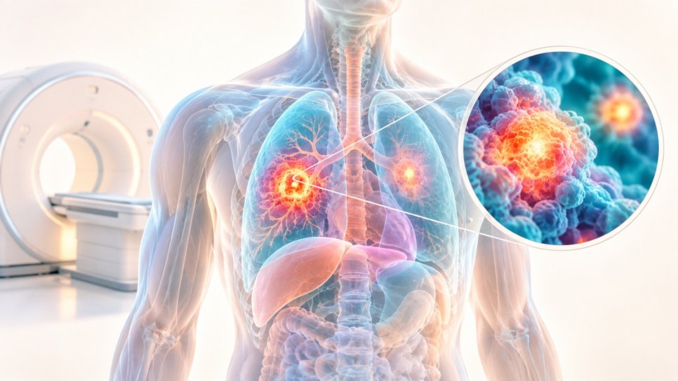
SUV stands for “standardized uptake value”. It summarizes in a single number how much of the radioactive tracer an area on the PET image has taken up. SUVmax is the highest SUV measured in the chosen area. The tracer most often used in PET/CT is FDG; it resembles glucose (sugar) and accumulates in tissues that use a lot of sugar.
| Topic | Detail |
|---|---|
| Calculation | The radioactivity concentration in the chosen area is divided by the injected dose divided by body weight |
| Other name | Dose uptake ratio (DUR) |
| Normalization | Body weight is used most often; values corrected for lean body mass or body surface area also exist |
| Which value appears on the report? | The highest (max), average (mean) or peak value |
| What does it show? | FDG uptake of the tissue, that is, its sugar use |
| What does it not show? | Whether the uptake comes from cancer, inflammation or infection; that distinction is made with the whole image and the clinical picture |
Source: Radiopaedia, “Standard uptake value” article.
Figure 1 · How is SUV calculated?
The two parts that make up the number, and the result
SUVmax, SUVmean and SUVpeak: which one appears on the report?
According to Radiopaedia, SUV can be reported for an area as the highest (max), average (mean) or peak value. The three give different numbers for the same lesion, so it matters which one is written on the report.
| Measure | What it describes | Point to watch |
|---|---|---|
| SUVmax | The highest value in the chosen area | Rests on a single point; can be affected by technical factors such as image noise, resolution and how the area was chosen |
| SUVmean | The average of the chosen area | Depends on how the borders of the area were drawn |
| SUVpeak | The average of a small zone around the most intense spot | Response criteria use the version corrected for lean body mass (SUL peak); we did not verify the size of the zone for this article |
When comparing two reports of the same patient, make sure the same measure was used.
What changes the SUV value?
SUV does not depend on the tissue alone. Factors related to the patient, to how the tracer is distributed in the body, and to the scanner can push the number up or down. A 2014 abstract from Emory University lists these groups:
| Group | Examples |
|---|---|
| Patient factors | Blood sugar after a meal, insulin, steroid use, acute illness |
| Conditions affecting distribution | Anemia, drugs that stimulate the bone marrow, excessive exercise, kidney failure, widespread swelling (anasarca), marked excess weight |
| Technical factors | Wrong units entered, different waiting times, the dose leaking outside the vein, the number of iterations and filters used in image reconstruction |
| Small lesion size | In small lesions, partial volume effect or breathing motion can make the value look lower than the truth |
Source: Plaxton et al., J Nucl Med 2014;55(Suppl 1):1356. The abstract concludes that nuclear medicine physicians need to know when SUV may be unreliable or underestimated.
This is why preparation before the scan matters. The text of the 2010 EANM FDG PET guideline says the following: fasting for at least 6 hours before the scan, the scan can go ahead if blood sugar is below 7 mmol/L (120 mg/dL), and the recommended interval between injection and image acquisition is 60 minutes. The same guideline states that differences in camera calibration, image reconstruction and data analysis can have an effect of more than 50% on the measured SUV.
Figure 2 · Standard scan sequence (guideline example)
Your center’s instructions may differ; follow your own center’s protocol
If SUV is high, is it cancer? Non-cancer causes
No, not on its own. FDG accumulates in any tissue that uses a lot of sugar: cancer cells are one of them, but infection and inflammation cells also use a lot of sugar. So on PET/CT a high SUV means “there is an active tissue”; to work out what that tissue is, the shape and location of the finding, the CT features, the patient’s history and, when needed, a biopsy are weighed together.
| Cause | What has been reported? |
|---|---|
| Infection | Mycobacterial, fungal and bacterial infections can give intense uptake. One review calls tuberculoma “one of the most well-known diseases that show intense FDG uptake” |
| Inflammation | Sarcoidosis and radiation pneumonitis can give intense uptake; in sarcoidosis it has been suggested that the intensity of uptake may reflect disease activity |
| Physiological | Brown fat has been reported as a false positive result in 2.3–4% of patients |
| Blood sugar | With high blood sugar, inflammatory cells use more glucose than at normal blood sugar, which can make inflamed areas more likely to be read as malignant |
| Around the tumor | About 24% of the FDG concentration in a tumor mass comes from non-tumor tissue |
Source: a lung FDG-PET review published on KoreaMed Synapse (article no. 1027719).
SUV examples reported in non-cancer diseases
Individual cases mentioned in the review; they are not typical values for these diseases.
In the same review, the SUV of the paragonimiasis case rose to 6.2 on the 2-hour image, an example showing that waiting time changes the number.
An SUVmax ≥2.5 threshold in a 32-patient lung nodule study
The threshold was chosen from the study’s own ROC curve. In percent.
Causes of the 5 benign nodules that stayed above the threshold: 2 histoplasmosis, 1 tuberculosis, 1 invasive aspergillosis, 1 nonspecific inflammation. The study is small (32 patients); its results cannot be carried over to every center.
For a lesion with a high SUV, the decision “cancer” or “not cancer” does not come from the number. The number shows where to look; the next step (comparison, further imaging, biopsy or follow-up) is chosen together with your physician. For what a biopsy is, see our article What is a biopsy? (in Turkish).
If SUV is low, does that mean no cancer?
No. According to the same review, tumors with low glycolytic activity are a well-known main cause of false negative results. In addition, in small lesions the number can come out lower than it should.
| Situation | What has been reported? |
|---|---|
| Bronchioloalveolar carcinoma | Lower FDG uptake than in adenocarcinomas has been reported; in one case the SUV is 1.7 (the term used by the review) |
| Carcinoid tumor | In one case the SUV was in the range 1.0–2.3; attributed to low metabolic activity |
| Mucinous tumors | Mucinous bronchioloalveolar carcinomas show significantly lower peak SUV than squamous cell carcinomas |
| Lesions smaller than 1 cm | Common PET systems have a resolution of about 1 cm, so high uptake may not be seen |
| Lesions smaller than 7 mm | Radiopaedia notes that sensitivity and specificity fall below this size |
Whether a lesion with a low SUV is followed or assessed by other methods is the physician’s decision; this table is not a diagnostic guide.
What does the “2.5 threshold” really tell us?
A frequently cited SUVmax number for lung nodules is 2.5. But it is not a universal boundary: Radiopaedia cites a range of 2.0–2.5 rather than a single number for separating benign from malignant, and the KoreaMed review says that 2.5 and above has been used as an indicator of malignancy and that this is controversial. In the small study in the Jornal Brasileiro de Pneumologia, 2.5 was chosen from the ROC curve of that study’s own patients.
The study enrolled 53 consecutive patients and excluded 21 (10 had not completed one year of follow-up, and 11 had a previous cancer). Of the remaining 32 patients, 14 had a malignant nodule. With a threshold of 2.5, 13 malignant nodules stayed above it and 1 malignant nodule stayed below it; of the 18 benign nodules, 5 were above the threshold.
Figure 3 · From the number on the report to a decision
SUVmax is a starting point, not a conclusion
SUV in treatment response: a short look at PERCIST
SUV is also used to compare PET scans before and after treatment. The criteria called PERCIST rely on the peak value (SUL peak) of SUV corrected for lean body mass. According to the summary we read, the most intense lesion before treatment is compared with the most intense lesion after treatment; they do not have to be the same lesion.
Figure 4 · Comparing two scans in PERCIST
Response is judged by the difference between two numbers, not by a single number
The page we read did not state the minimum baseline uptake, glucose and waiting time conditions, so we have not written them here. You can ask your physician which criteria were used in your report.
What to watch for when comparing two reports
The EANM guideline recommends the same waiting time in repeat scans (with a tolerance of ±5 minutes), the same PET or PET/CT system, and the same image acquisition and reconstruction settings. The SUVs of two reports scanned at different centers or on different scanners may therefore not be directly comparable.
- Check which measure (max, mean, peak) the report gives for SUV.
- Ask whether the earlier scan was done at the same center and on the same scanner.
- Find out whether the fasting time and blood sugar before the scan were recorded.
- Ask whether the time from injection to image acquisition was similar in the two scans.
- Tell the physician about steroids, drugs that stimulate the bone marrow, or a recent infection.
- Follow the center’s preparation instructions before the scan in full.
Practical tips and common misconceptions
- “A high SUVmax is definitely cancer.” No. Infection, inflammation and brown fat can also give high uptake.
- “A low SUVmax means there is no cancer.” No. Some tumors, such as carcinoid and mucinous tumors, and very small lesions can give low values.
- “Above 2.5 is malignant, below is benign.” No. There is no single universal threshold; sources cite a range of 2.0–2.5 and its use is controversial.
- “SUVs from two different centers can be compared directly.” Not always. Differences in scanner, image reconstruction and analysis can change the measured value by more than 50%.
- “Eating or drinking before the scan doesn’t matter.” No. Blood sugar after a meal is among the patient factors that affect SUV; follow the center’s preparation instructions.
Questions to ask your physician
- Which lesion is this SUVmax given for, and how was it measured?
- Were my blood sugar and waiting time before the scan recorded?
- Could this uptake have a non-cancer explanation (infection, inflammation, brown fat)?
- If the lesion is small, could the value have been measured lower than the truth?
- Was I scanned on the same scanner and with the same protocol as before?
- What should the next step be: a repeat scan, other imaging, a biopsy or follow-up?
For this article we did not check the preparation instructions of centers in Türkiye, differences in scanners and software, report formats or reimbursement conditions; we made no assumptions on these. For when PET/CT is used, see our article What is PET/CT? (in Turkish), and for biopsy, What is a biopsy? (in Turkish).
DROZDOGAN Academy Assessment
SUVmax is the number people wonder about most on PET/CT reports, and patients often look at it like a “score”. In reality this number is a measure of how intense the uptake is, and it gains meaning only together with the images, the clinical information and, when needed, a tissue diagnosis.
⚡ Why it matters
Over-reading or under-reading a single number causes problems in both directions: a value that is high for a non-cancer reason can lead to needless worry, and a low value can lead to false reassurance. This assessment is our own.
✅ What is strong
The definition of SUV, how it is calculated, the groups of factors that affect it, scan preparation and the principle of consistency in repeat scans appear consistently in more than one source. We have concrete examples of non-cancer causes and of low-uptake tumors, and the numerical results of one study.
⚠️ What is weak
Some of the numerical examples rest on single cases and on a 32-patient lung nodule study. The EANM text was read from the 2010 version; a newer version exists and its numbers may differ. The PERCIST information was taken from a textbook page, not from the original article.
🩺 What it means in practice for patients and physicians
When you receive the report, instead of focusing on the number alone, read the nuclear medicine physician’s interpretation and the whole set of images together. Ask your physician whether you can choose the same center and protocol for comparison with earlier scans.
❓ Open questions
The sources of this article cannot answer what “normal” and “suspicious” SUV ranges are for different cancer types, how large the difference between scanners is in practice, or the details of the current guideline.
Bottom line: SUVmax is a useful number that shows how intense the uptake is; on its own it does not show whether cancer is present or absent. Consult the physician who will assess the number on your report together with the images; do not draw conclusions on your own.
Frequently asked questions
- What SUVmax value means cancer?
- There is no single number like that. For lung nodules, a range of 2.0–2.5 and a figure of 2.5 are often cited, but their use is controversial and benign lesions are also found above the threshold.
- My SUVmax is high, do I have cancer?
- That cannot be concluded from this number. Infection, inflammation and brown fat can also give high uptake. Talk to the physician who assesses the whole set of images.
- If SUVmax is low, am I safe?
- That cannot be said from the number alone either. Some tumors give low uptake, and in lesions smaller than 1 cm the value may be measured lower than the truth.
- What is the difference between SUVmax and SUVmean?
- SUVmax is the highest value in the chosen area; SUVmean is the average of the chosen area. They give different numbers for the same lesion.
- Why is fasting needed before the scan?
- Blood sugar after a meal is among the factors that affect SUV. The 2010 EANM guideline text cites fasting for at least 6 hours and blood sugar below 7 mmol/L; your own center’s instructions may differ.
- Can I compare SUV values from two different centers?
- Not always. Differences in scanner calibration, image reconstruction and analysis can change the measured value by more than 50%; the guideline recommends the same scanner and settings for repeat scans.
- What is PERCIST?
- A set of criteria that classifies response by comparing SUL peak values on PET scans before and after treatment. For a partial response, a reduction of more than 30% and of more than 0.8 units is required.
Glossary
- PET/CT
- Imaging that combines PET, which shows metabolic activity, with computed tomography, which shows anatomical detail.
- FDG
- A radioactively labeled sugar that resembles glucose; the tracer most often used in PET/CT.
- SUV
- Standardized uptake value; a measure of how much of the tracer an area has taken up.
- SUVmax
- The highest SUV value in the chosen area.
- SUVmean
- The average SUV value of the chosen area.
- SUVpeak / SUL peak
- The average of a small zone around the most intense spot; SUL is the version corrected for lean body mass.
- False positive
- A result the test calls “suspicious” that is not actually cancer.
- False negative
- A result the test calls “normal” although a tumor is actually present.
- ROC curve
- A graph showing a test’s sensitivity and specificity at different thresholds; used to choose a threshold.
- Sensitivity and specificity
- Sensitivity is the rate at which people with the disease are detected; specificity is the rate at which people without the disease are correctly ruled out.
Verified: the definition and formula of SUV, the normalization options, the max/mean/peak distinction, the factors, the 2.0–2.5 range and the 7 mm statement were read from the Radiopaedia article; the factor groups from the Emory University J Nucl Med 2014 abstract; fasting, blood sugar, waiting time, repeat scans and the 50% statement from the 2010 text of the EANM FDG PET guideline. The PERCIST information was taken from the web page of a nuclear medicine textbook. The non-cancer causes and low-uptake tumors were read from a lung FDG-PET review on KoreaMed Synapse; the 32-patient study from the article in the Jornal Brasileiro de Pneumologia. The abstract of that study says “one false positive”, but its results section and the specificity of 72.2% fit 5 false positives; we relied on the results section. Only noted: version 2.0 (2015) of the EANM guideline exists; we did not read it, so the numbers there may differ. The title and author details of the review could not be taken from the page. We could not read the original PERCIST article because the PMC and PubMed pages were not accessible. Not verified and therefore not written: “normal” SUV ranges and threshold values for different cancer types; the visual scales used in lymphoma; the numerical size of differences between scanners; the practices of centers in Türkiye; overlap with articles on drozdogan.com (not checked). Assessments and opinions belong to this article. This article is not medical advice; no diagnosis should be made and no treatment decision taken on its basis.
Who is this article for? Patients who see an SUV value on a PET/CT report, their relatives and health professionals interested in the topic. This article is not medical advice; only the physicians who follow you can determine what your report means.
References
- Radiopaedia. Standard uptake value. radiopaedia.org
- Plaxton N, Moncayo V, Barron B, Halkar R. Factors that influence standard uptake values in FDG PET/CT. J Nucl Med 2014;55(Suppl 1):1356. jnm.snmjournals.org
- EANM. FDG PET and PET/CT procedure guideline, version 1.0 (2010). link.springer.com
- PERCIST criteria (textbook page). numebook.uchile.cl
- KoreaMed Synapse. Review of FDG-PET in the lung, article no. 1027719. synapse.koreamed.org
- Jornal Brasileiro de Pneumologia. FDG-PET evaluation of the solitary pulmonary nodule (53 consecutive patients). jornaldepneumologia.com.br
Was this article helpful?
Your feedback helps us improve our content.



