
A Genius Who Died at Thirty: Xavier Bichat and Six Hundred Autopsies in One Year

In this article
The Moment Pathology’s Resolution Moved from Organ to Tissue (1800-1802)
On July 22, 1802, a small funeral gathering assembled in the courtyard of Hôtel-Dieu Hospital in Paris. It was raining. Young medical students and a few colleagues stood beside the coffin; among them, a man stood a little apart from the crowd behind him — a man who would later become Emperor Napoleon’s personal surgeon and one day operate on Frances Burney, Baron Dominique-Jean Larrey — quietly weeping. The person lying in the coffin was a physician aged thirty . Two weeks earlier, on July 8, 1802, he had fallen down the marble stairs of Hôtel-Dieu and struck his head. He suffered a week of intense fever and delirium, then lost consciousness and died on the morning of July 22. His name was Marie François Xavier Bichat. He left behind four books, approximately five thousand autopsies (an estimated 600 in the last year alone), a medical revolution, and dozens of students. When Napoleon received news of his death, he had the following sentence written: “We have lost a man who, had he lived a few more years, could have changed the face of medicine single-handedly.” He also ordered the late Bichat’s bust to be placed in the courtyard of Hôtel-Dieu beside that of his teacher, Pierre-Joseph Desault. What Bichat accomplished in this brief life was to take the revolution in pathological anatomy that Morgagni began in 1761, discussed in yesterday’s article, one step further: disease resides not in an organ, but in the tissue within that organ. The description and classification of 21 tissue types is the birth certificate of modern histology. There is scarcely another figure in medical history who initiated such a profound transformation in so short a life.
The Thirty Years of a Physician Born on November 14, 1771, and Deceased on July 22, 1802
Bichat was born on November 14, 1771, in the village of Thorette in the Ain region of eastern France. His father, Jean-Baptiste Bichat, was both a physician and his first anatomy teacher within the family — just as Frances Burney’s father was a musicologist, Bichat’s father introduced the young boy to medicine early. The family moved to Poncin in the Rhine-Alps region, where young Xavier received his first education.
In 1782, aged 11, he enrolled at the college in Nantua, and in 1790 studied philosophy and rhetoric at the Saint-Irénée seminary in Lyon. Around the same time, two surgeons in Lyon, Marc-Antoine Petit and Jean-Marie Viricel, took responsibility for his education in anatomy and surgery. But the decision that transformed Bichat’s life came in 1793: he moved from Lyon to Paris. France was in revolutionary chaos, the Terror had not yet ended, but Paris remained the scientific center of Europe.
In Paris, Bichat became an assistant to the most brilliant surgeon of his time, Pierre-Joseph Desault(1738-1795). Desault was chief surgeon at Hôtel-Dieu Hospital; he had pioneered orthopedic and trauma surgery and led the largest school of surgical observation in Paris. At his side for two years, Bichat experienced the most intensive practice of anatomy, clinical observation, and pathological anatomy in Paris. When Desault died suddenly in 1795, completing his teacher’s unfinished work fell to Bichat: publishing Desault’s surgical memoirs, organizing the remaining files, and running his school. This was an overwhelming burden for a physician of only 24.
From 1797 onward, Bichat began teaching independently. Young physicians in Paris flocked to him; his lectures became so popular that the lecture theaters of Hôtel-Dieu were insufficient, and he organized private classes in his own home. In 1799, again on his own initiative, he founded the Société Médicale d'Émulation— a society of young physicians seeking to raise professional standards. His principal field of work was defined: reestablishing the link between anatomy and pathology.
The Extraordinary Working Pace of a Pathologist Without a Microscope
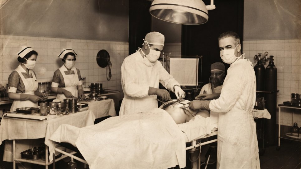
From 1799 onward, Bichat abandoned surgery and devoted himself full-time to anatomical research. The morgue at Hôtel-Dieu was his laboratory. A number that is hard for the modern reader to believe: approximately 600 autopsies in a single year. This means the dissection of an average of two human bodies every day — clinical lectures in the mornings, autopsies in the afternoons, writing and reading in the evenings. Three factors lay behind this pace: continual deaths in the crowded hospitals of post-Revolutionary Paris, Bichat’s plainly extraordinary capacity for work, and an obsession lodged in his mind — where does disease reside?
Bichat knew Morgagni’s answer from 1761: disease resides in the organ. But Bichat saw that this answer was insufficient, because different parts of the same organ were susceptible to different diseases. Stomach cancer often began in the pyloric region but rarely in the stomach body; liver cancer appeared either as nodules distributed throughout the entire organ or in a form surrounding the bile ducts. Pulmonary tuberculosis affected the alveoli, whereas lung cancer settled in the bronchial tree. Thus, the question which part of the organ is diseased? was more precise than Morgagni’s question — and answering it required looking below the level of the organ.
This was Bichat’s real contribution: tissue is the building block of the organ. An organ is a combination of different tissues in a particular architecture; the same tissue type can be found in different organs, and a disease is generally linked to tissue rather than to the organ. For example, “disease of serous membranes” — peritoneum, pleura, pericardium — was the manifestation of the same tissue in different organs. Tuberculosis was a disease of serous membranes (pericarditis, peritonitis, and pleuritis all together); pneumonia was a disease of alveolar tissue, and hepatitis of the hepatic parenchyma. This is a fundamental principle that Virchow (Day 87) and modern molecular pathology would later also accept in versions with finer resolution: the same tissue type carries the same pattern of disease.
Histology Classified Without a Microscope
Between 1800-1801, Bichat published three fundamental works: Traité des membranes en général et de diverses membranes en particulier (A General Treatise on Membranes and on Various Membranes, 1800), Recherches physiologiques sur la vie et la mort (Physiological Research on Life and Death, 1800), and his masterpiece, Anatomie Générale appliquée à la physiologie et à la médecine (General Anatomy Applied to Physiology and Medicine, 1801). The latter was an immense four-volume work in which Bichat systematically classified the 21 tissue types he distinguished with the naked eye, boiling, acid/base reactions, and mechanical dissection.
BICHAT’S 21 TISSUE TYPES — FROM ANATOMIE GÉNÉRALE, 1801
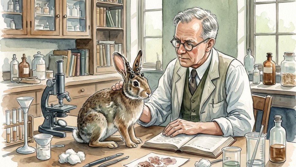
What is surprising about this list from a modern perspective is that Bichat used no microscope at all. In fact, Bichat did not trust the microscope — he regarded it as an uncontrolled, misleading instrument. In the early 1800s, microscopic instruments had not yet overcome the problem of “chromatic aberration” (color distortion), which produced colored rings and blurred images. Adequate optical correction would have to await Joseph Jackson Lister’s invention of the achromatic objective in the 1830s (he was the father of Joseph Lister, the father of surgical antisepsis). Bichat’s statement “I do not trust the microscope” was scientifically reasonable in 1800. The instrument itself was not yet reliable for pathological anatomy.
The methods Bichat used to distinguish tissues can today be seen as the ancestor of histochemistry : boiling (a change in consistency through protein denaturation), treatment with acid (collagen and elastin reactions), treatment with a base, drying, roasting, and solvents such as ether and alcohol. Each tissue responded differently to these stimuli, and Bichat systematically recorded the differences. Thus, his description of 21 tissue types is a taxonomy based on systematic observation of chemical and mechanical properties .
The Impact of Tissue Pathology on Oncology
Bichat’s work did not focus directly on oncology; he was a physiologist and anatomist interested in a general theory of disease. But the framework he described laid the foundations for the following century’s understanding of cancer. Several important consequences:
First, questioning the tumor’s tissue of origin. When Morgagni spoke of “stomach cancer,” he referred to an organ; after Bichat, physicians began asking, “Which tissue of the stomach is diseased?” This is the philosophical ancestor of Wilhelm Waldeyer’s distinction in 1867 between carcinoma (of epithelial origin) and sarcoma (of mesenchymal origin), discussed on Day 90. The idea that carcinoma arises from an epithelial cell extends the placement of epithelial-like structures numbered 10-15 among Bichat’s 21 tissues in a separate category.
Second, interpreting the tumor’s anatomical pattern of spread through a tissue-centered perspective. Bichat observed a shared pattern of disease in serous membranes (peritoneum, pleura, pericardium): peritoneal carcinomatosis, pleural metastasis, and pericardial effusion — all are similar cancer reactions of serous tissue. Modern oncology still uses this concept today: “peritoneal spread, pleural effusion” and similar descriptions carry the categories Bichat established 200 years ago.
Third, the theoretical foundation that led to the concept of histopathological diagnosis. When the microscope entered pathology after the 1830s — following Joseph Jackson Lister’s achromatic objective — pathologists sought the microscopic counterpart of each of Bichat’s 21 tissues. When Johannes Müller published microscopic descriptions of tumors in 1838 (Day 81), the classification framework he used was largely based on Bichat’s tissue categories. Modern histopathological tumor classification (the WHO tumor atlas) is still built on Bichat’s concept of tissue.
AN ASIDE: THE “VITALISM” DEBATE
Alongside his tissue doctrine, Bichat also participated in the intellectual debates of his time. In Recherches physiologiques sur la vie et la mort, he argued that life rests on “two separate forces”: animal life (relations with the outside world: sensation, muscle, will) and organic life (internal continuity: digestion, circulation, reproduction, autonomic functions). These two forces were expressed in different tissue types. The term “autonomic nervous system”, an important concept in modern biology, is a direct extension of Bichat’s category of “organic life.” Bichat is also known as one of the last great theorists of vitalism— the view that life is governed by a “vital force” distinct from chemistry and physics alone. Cell theory (1839, Schwann-Schleiden) and the subsequent development of biochemistry would leave vitalism as a historical transitional stage; but Bichat’s tissue doctrine survived even when the rejection of vitalist philosophy followed him.
The Unexpected End of Science
THE STAIRS OF HÔTEL-DIEU
On July 8, 1802, after finishing his lecture at Hôtel-Dieu Hospital, Bichat was descending the stairs — according to modern historians, probably because of fatigue or a mild headache that day — when he lost his balance and fell. He struck his head on a marble step. For the first few days, there was no obvious problem apart from mild dizziness, and he did not postpone his lectures. But around July 14, high fever and headaches began. On July 18, loss of consciousness. On the morning of July 22, 1802, he died in Paris, aged 30.
The most likely diagnosis from a modern reading: chronic subdural hematoma associated with head trauma + secondary infection (probably meningoencephalitis). But it remained a mystery to the medicine of the time; some contemporaries attributed it to “overwork”, others to “tuberculosis from the bad air in the autopsy room”. A year before his death, in the autumn of 1801, he had suffered an episode of hemoptysis (bloody saliva) — so he may have had active or ongoing pulmonary tuberculosis, which may also have worsened his complications after the head injury.
Bichat had published four works before his death. Of his fifth work, Anatomie descriptive(Descriptive Anatomy), he was able to complete only the first two volumes; his students compiled the remaining two from his notes and finished them in 1803. When Napoleon received news of his death, he had a bust of Bichat erected in the courtyard of Hôtel-Dieu Hospital — beside the bust of his teacher Desault. The two busts still stand there today, side by side in the courtyard of the modern Hôpital Hôtel-Dieu. Stone portraits of two men whose lives burned like a fire, in front of the oldest hospital in Paris.
224 Years from Tissue Doctrine to Modern Histopathology
📖 BICHAT’S LINK IN PATHOLOGY’S CHAIN OF RESOLUTION
The 21 tissue types Bichat defined in 1800-1801 are handled in a different taxonomy in modern histology — essentially four main tissue groups: epithelial tissue, connective tissue (including bone and cartilage), muscle tissue, and nervous tissue. Modern classification rests on microscopic observation and cellular structure; Bichat’s naked-eye classification is coarser but fundamentally built on the same logic. His category of “serous membrane” is read in modern histology as a single epithelial type (mesothelium); the category of “mucous membrane” encompasses different epithelial types (columnar, stratified squamous, etc.). From a modern perspective, some of Bichat’s categories merge and others split apart; but the basic idea survives: tissue, not organ — the taxonomic unit of disease.
Even today’s molecular oncology carries this principle. A single lung cancer is not simply “lung cancer”: squamous cell carcinoma, adenocarcinoma, small-cell carcinoma, and large-cell carcinoma are each tumors of different tissue origins (bronchial epithelium vs alveolar epithelium vs neuroendocrine cells), requiring different treatment profiles. This distinction is a modern extension of Bichat’s tissue distinction from 1800.
The Same City, the Same Decade, Two Different Lives
Here, an interesting historical coincidence bringing together the last three days of the series deserves mention. Bichat of our Day 73 died in the Paris where our Day 71’s Frances Burneyunderwent surgery (her Paris mastectomy in 1811), exactly nine years earlier . Burney’s period living in Paris, 1801-1812, coincides with the time immediately after Bichat’s death. Baron Larrey and Antoine Dubois, who operated on Burney, were Bichat’s contemporaries — Larrey in particular may have been among those who attended Bichat’s funeral (this is not certain, but is likely given the period and their professional relationship). Dubois was Bichat’s contemporary at Hôtel-Dieu, and Bichat’s clinical lectures were a reference point for all the leading surgeons in Paris.
Burney could not say in her letter what the tissue removed in her 1811 operation was — because pathology at the time was not yet mature. But if the same tumor had been in Bichat’s hands, if he had been able to open and examine it — the structural similarities between a scirrhous stomach tumor and a scirrhous breast tumor, the interaction between fibrous and glandular tissue, the reaction of the surrounding lymphatic tissue — Bichat would probably have classified it within his own framework of 21 tissues. But he had no time to do so. Nine years before Burney’s 1811 operation, Bichat had died at 30; nevertheless, some of the intellectual atmosphere surrounding the physicians who operated on Burney was indebted to Bichat’s tissue doctrine.
A BRIEF FACT-CHECK NOTE
- Bichat’s birth and death: November 14, 1771, Thorette (Ain, France) — July 22, 1802, Paris; aged 30
- His father: Jean-Baptiste Bichat, physician and first anatomy teacher
- Education: Nantua college in 1782, Lyon (Saint-Irénée) in 1790-1792 (philosophy + rhetoric + surgery); his teachers were Marc-Antoine Petit (not to be confused with Jean-Louis Petit) and Jean-Marie Viricel
- Arrival in Paris: 1793, the end of the Revolution’s Terror period
- His teacher in Paris: Pierre-Joseph Desault (1738-1795), chief surgeon at Hôtel-Dieu; after Desault’s death, Bichat took over his school as his assistant
- Founding of the Société Médicale d'Émulation: 1796 (some sources give 1799); a society founded by Bichat for young physicians to establish professional standards
- Abandoning surgery: Full-time anatomical research from 1799 onward
- Annual number of autopsies: Approximately 600/year in the last three years of his life (an estimated total of 5.000-6.000 autopsies)
- He did not use a microscope: The problem of chromatic aberration had not been solved; he found the instrument unreliable
- Principal works: Traité des membranes (1800), Recherches physiologiques sur la vie et la mort (1800), Anatomie générale (1801, 4 volumes), Anatomie descriptive (1801-1803, 5 volumes; the last two compiled by his students after his death)
- 21 tissue types: The principal taxonomic contribution of Anatomie générale
- Cause of death: A fall on the stairs of Hôtel-Dieu on July 8, 1802; from a modern reading, head trauma + possible secondary infection and/or complications; the official diagnosis was not certain at the time; an additional possibility: active pulmonary tuberculosis (he had an episode of hemoptysis in the autumn of 1801)
- Napoleon’s decision: He had busts of Bichat and his teacher Desault erected in the courtyard of Hôtel-Dieu
René Laennec and the Invention of the Stethoscope — 1816, Paris
Fourteen years after Bichat’s death, in the same Paris, a thirty-five-year-old physician found it embarrassing to place his ear against his patient’s chest, and listened to the heart through a rolled paper tube in front of him. He heard the sound with surprising clarity. His name was René Laennec; what he invented that day became the stethoscope that every physician carries around the neck today. Tomorrow, we will tell the story of lung cancer becoming, for the first time in history, a disease a physician “heard” — and the tragic cycle of Laennec himself also dying of tuberculosis.
Author’s note: The Bichat quotation in this article was adapted into Turkish from the introduction to the Anatomie Générale 1801 edition; it is a conceptual summary of the original French text, not a literal translation. Bichat’s list of 21 tissue types does not correspond exactly to modern histological classification; some are divided into different epithelial or connective-tissue subtypes in a modern reading, while others are merged. The list presented in this article gives Turkish equivalents of Bichat’s original categories. The exact cause of Bichat’s death has not been historically established; “a fall on the Hôtel-Dieu stairs + head trauma” is the prevailing view, but active pulmonary tuberculosis, meningoencephalitis, or other complications are also possibilities mentioned in sources of the period.
References and Further Reading
- Bichat MFX. Traité des membranes en général et de diverses membranes en particulier. Paris: Richard, Caille et Ravier, 1800.
- Bichat MFX. Recherches physiologiques sur la vie et la mort. Paris: Brosson, Gabon et Cie, 1800.
- Bichat MFX. Anatomie générale appliquée à la physiologie et à la médecine. 4 volumes. Paris: Brosson et Gabon, 1801.
- Bichat MFX. Anatomie descriptive. 5 volumes. Paris: Brosson et Gabon, 1801-1803. (The last two volumes were completed by his students after his death.)
- Perdicoyianni-Paleologou H. Xavier Bichat and the renovation of the pathological anatomy. Folia Medica 2022;64(2):203-211. (A modern reference article on biography and the history of science.)
- Huard P, Imbault-Huart MJ. Bichat, anatomiste et physiologiste. Paris: Vrin, 1971. (The standard French-language academic biography of Bichat.)
- Maulitz RC. Morbid Appearances: The Anatomy of Pathology in the Early Nineteenth Century. Cambridge: Cambridge University Press, 1987. (A classic medical history source on Bichat and his era.)
- Albury WR. Bichat, Xavier (1771-1802). New Dictionary of Scientific Biography, ed. Noretta Koertge, Detroit: Charles Scribner's Sons, 2008.
- Long ER. A History of Pathology. New York: Dover Publications, 1965. (The historical framework of the Morgagni-Bichat-Virchow chain of pathological anatomy.)
- Mukherjee S. The Emperor of All Maladies: A Biography of Cancer. New York: Scribner, 2010. (The long-term impact of Bichat’s tissue doctrine on modern oncology.)
- Encyclopedia Britannica. Marie-François-Xavier Bichat: cellular anatomy, pathology, physiology. Accessed: 2025.
- Bichat MFX. A Treatise on the Membranes in General, and on Different Membranes in Particular. Translated by John Coffin. Boston: Cummings & Hilliard, 1813. (The first English translation of Traité des membranes.)
This article is Day 73 of the History of Cancer in 365 Days Series. Pathology’s chain of resolution extends from Bichat’s tissue doctrine to microscopic pathology, cellular pathology, and molecular pathology; the subsequent links in this chain will be discussed through Müller (Day 81), Schwann-Schleiden’s cell theory (Day 84), Virchow’s Cellularpathologie (Day 87), and Waldeyer’s carcinoma-sarcoma distinction (Day 90). The article was prepared from a historical and popular perspective; it contains no medical treatment guidance.
Was this article helpful?
Your feedback helps us improve our content.


Reader comments
Share your comment
Your name and comment are published only after administrator approval. Your email address is never shown to visitors.
Be the first to comment.