Seeing Inside the Body Without Cutting: Damadian, Lauterbur, Mansfield and the 1977 of Magnetic Resonance
In this article
Seeing Inside the Body Without Cutting: Damadian, Lauterbur, Mansfield and the 1977 of Magnetic Resonance
On Day 162 we watched a drug put a lock on a hormone receptor. Today we move to another scene from the same year, 1977; as I promised in yesterday's tomorrow box, I will first separate out what 1977 is actually the date of. On Day 154, Hounsfield's computed tomography opened a way to look inside the body with X-rays; on Days 92 and 95 we saw the cost of seeing, and of treating, with radiation. Today's story is about a method that tries to look inside the body without ionizing radiation, how it grew out of three people's three different contributions, and the years-long argument over who owns them.
A section that took five hours
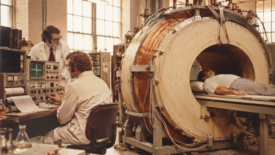
According to an account in Smithsonian magazine, Raymond Damadian's team began the first test on 11 May 1977 with a homemade machine they called "Indomitable". Its magnet was made by winding about 30 miles (about 48 kilometres) of niobium-titanium wire on a cylinder; it was about 53 inches across and weighed about 1.5 tons, and its leaking liquid-helium cooling cost about 2,000 dollars a week. The first subject was Damadian himself, but no signal was obtained; the team put this down to body fat insulating the signal. They moved on to a leaner graduate student, Larry Minkoff.
The same account says the first human image was obtained on 3 July 1977, nearly five hours into the test. The company Damadian founded, Fonar, gives the completion time in its own timeline as 04:45 on 3 July 1977. The image was a crude two-dimensional section of Minkoff's chest, including the heart and lungs; it was built first with coloured pencils and then by computer from 106 data points. An antenna coil of cardboard, capacitors and copper wire, made by Mike Goldsmith, was worn by the subject. Two sources conflict on Minkoff's status: Smithsonian says "graduate student", Fonar says "postdoctoral fellow".
Which was the "first human image"? It depends on the definition
"The first human MRI image" does not point to a single event. According to Mansfield's Nobel biography, line-scan images of a finger made in Nottingham (with scan times of 15-23 minutes) were presented at a Medical Research Council (MRC) meeting in 1976 and persuaded the council to support a whole-body MRI project; the paper in the British Journal of Radiology is dated March 1977 (Mansfield and Maudsley, volume 50, pages 188-194). So an image of a human body part comes before Damadian's chest section. Damadian's 3 July 1977 image is recorded as the first whole-body attempt that produced a chest section of a living person. In April 1978 Mansfield imaged his own abdomen in a 50-minute scan (Nobel biography); the image of a living human forearm by Hinshaw and colleagues is dated January 1979 (Br J Radiol 52:36-43).
And the link to cancer? The 1977 chest section did not image a tumour; no account mentions one. According to the University of Aberdeen's own news item, the first whole-body MRI scan of a patient was made on 28 August 1980: the target was tumours in the liver of an elderly man with advanced cancer from Fraserburgh, and the scan was performed by the radiologist Francis Smith with the scanner developed by Hutchison, Redpath, Edelstein and Johnson in the medical physics department headed by John Mallard. The page does not report the findings of the scan. So 1977 is not the year of the first MRI image (1973), of the first clinical cancer scan (1980) or of clinical use: it is the year of the first human images.
How does MRI make an image?
In a strong magnetic field, some of the hydrogen nuclei in tissue (mostly in water and fat) line up with the field. A brief radio pulse disturbs this alignment; as the nuclei return to their former state, the speed of the return is described by two times that differ between tissues (the T1 and T2 relaxation times). An image needs two things: telling these different signals apart, and knowing where in the body each signal comes from. The second is done by making the field vary from place to place, that is, with a gradient, so that position is encoded in the frequency of the signal. This is a simplified account that gives the main line.
Damadian's idea: does a tumour "relax" differently?
Damadian's contribution begins with the paper "Tumor detection by nuclear magnetic resonance", published in Science on 19 March 1971 (volume 171, pages 1151-1153; the date is from Fonar's timeline). According to the Smithsonian account it rested on measurements of rat liver samples in 1970; the idea was that tumour tissue holds more water, and therefore more hydrogen, and that the signal lasts longer. The site mriquestions reports that the paper described longer T1 and T2 in several cancers than in normal tissue. On 17 March 1972 Damadian filed a patent application for a method of detecting cancer in tissue; the patent (US 3,789,832) was granted on 5 February 1974. The site mriquestions says the application did not include how an image would be formed; later court rulings came out in Damadian's favour (see below). I have not read the patent text.
Part of the idea turned out to be wrong. According to a column by Peter Rinck (himself a figure in the field, so a secondary source), Hollis and colleagues showed within a few months that tumour T1 is not necessarily longer than in other diseased or normal tissues (Cancer Research 1973;33:2156-2160); in 1985 Rinck and colleagues concluded that in vivo T2 measurements could not be used to detect, characterise or type cancer (Radiology 157:103-106). But according to the same column, the difference in relaxation times proved useful not for tissue diagnosis but for image contrast: T2-weighted spin-echo images made many diseases easy to see, and today one of the main MRI techniques in cancer diagnosis and follow-up rests on relaxation contrast. Damadian's hypothesis was wrong as a diagnostic tool; in another form, as image contrast, it stayed right.
Lauterbur and Mansfield: encoding position
According to Rinck's history article, Paul Lauterbur wrote a handwritten, countersigned draft on 2 September 1971 ("Spatially Resolved Nuclear Magnetic Resonance Experiments"); Nature rejected his first submission in late 1972. The paper appeared on 16 March 1973: "Image formation by induced local interactions: examples employing nuclear magnetic resonance" (Nature 242:190-191). According to mriquestions he used a 1.4 tesla magnet, four projections and back-projection. According to the Nobel text, Lauterbur showed that adding a gradient to the magnetic field could produce a two-dimensional image, demonstrating it in 1973 with tubes of ordinary water surrounded by heavy water. The same article says he urged his university to file a patent, but no application was made.
According to his Nobel biography, Peter Mansfield proposed NMR diffraction in solids after a tea-room conversation with Grannell and Garroway in Nottingham in 1972; the work of Mansfield and Grannell was presented in Krakow in September 1973 and published in the Journal of Physics C in November 1973. According to the Nobel text, Mansfield extended the use of gradients, showed how the signals could be analysed mathematically, and developed the very fast imaging method (echo-planar imaging) that became clinically useful about ten years later.
Cost and limits
The cost of the error was more than a scientific mistake. According to the Smithsonian account, on 20 July 1977 Damadian announced in a press release that a technique for non-surgical cancer detection had been "perfected". What had been obtained was a section of Minkoff's chest; the accounts do not say that a tumour was imaged. Rinck's article says Damadian withdrew his claim that he could detect cancer non-invasively at a press conference in 1977; I could not confirm this with another source. The first human scans still took hours (about five hours in 1977, 50 minutes in 1978 for Mansfield's abdomen) and the images were crude. The first machine was handmade, and its helium leak cost about 2,000 dollars a week.
The question of ownership also carried a cost. Damadian's company Fonar won its patent case against General Electric in 1997; Smithsonian gives 128 million dollars in damages, AuntMinnie a 62 million dollar ruling (I have put the conflict in the fact-check box). In this article I give no verified figure for MRI's safety restrictions, its cost or inequality of access for patients; these are the subject of a separate article.
What remained
- Encoding position with a gradient and building the image mathematically (Lauterbur, Mansfield)
- Using differences in relaxation between tissues as image contrast
- Very fast imaging (Mansfield's echo-planar method, clinically useful about ten years later)
What fell or was limited
- Diagnosing cancer from relaxation times alone (Damadian 1971; Rinck 1985)
- The 20 July 1977 announcement that non-surgical cancer detection had been "perfected"
- Telling the story of MRI as one person's invention
The first dates of MRI: 1971-2003. The axis runs from 1960 to 2005. Green markers are dates confirmed in more than one record; blue markers rest on a single source. Dates known only by year are drawn at mid-year.
The prize and the objection
On 6 October 2003 the Nobel Assembly awarded the Prize in Physiology or Medicine to Paul Lauterbur and Peter Mansfield "for their discoveries concerning magnetic resonance imaging"; the published text does not mention Damadian. A few days later, on 9-10 October 2003, Damadian and Fonar ran full-page advertisements headed "The Shameful Wrong That Must Be Righted" in the New York Times, the Washington Post and the Los Angeles Times; a second advertisement signed "Friends of Raymond Damadian" followed on 20 October (AuntMinnie). He told Nature: "If I had never been born, there would never be MRI today" (Nature, 16 October 2003). Damadian and Lauterbur had shared the National Medal of Technology in 1988 (Smithsonian).
We know the committee's grounds from the published text: image formation (the gradient, the mathematical analysis, the fast method). We do not know why Damadian was left out. According to AuntMinnie the committee does not comment on those not chosen and keeps its deliberations secret for 50 years; the historian Robert Marc Friedman is reported to think that Damadian's reputation may have played a part. That is a guess. People around Damadian also say that Lauterbur deliberately never cited Damadian (Minkoff's claim; one of the parties). Two separate questions are being mixed here: who first said that differences in the relaxation of tissues could be a sign of cancer (Damadian 1971, although that idea rests on the work of Odeblad and Lindström on relaxation times in the 1950s), and who first put forward an imaging method that encodes position (Lauterbur 1973; Mansfield-Grannell 1973)? Separating them does not mean saying who is right: the Nobel text rewarded the second question.
No verified local link
I could not find a verified source on in which year, and to which hospital, the first MRI scanner came to Türkiye, and I am not drawing a link. A Turkish journal article I looked at says only that the medical use of MRI began in the early 1970s with the experiments of Damadian and Hollis; it contains no information on Turkish history. I can write a general inference: for the number of scanners, waiting times and access for a method that is now routine in cancer diagnosis and follow-up, national registries have to be consulted; there is no such figure in this article.
From 1971 to today
What was confirmed, what stayed uncertain
- Day 158: Köhler and Milstein has still not been written; the promise in the tomorrow box of Day 157 was not kept.
- What 1977 is the date of: of the first human images: the Mansfield-Maudsley finger paper (March; presented at an MRC meeting in 1976), Damadian's chest section (3 July) and Damadian's press release (20 July). Not the first MRI image (1973), not the first patient scan (1980). "First human image" changes with the definition: for a body part (a finger) it is Mansfield, for a whole-body section it is Damadian.
- Time of day on 3 July 1977: 04:45 in Fonar's own timeline; Smithsonian says "nearly five hours into the test". The two agree, but I cannot say they come from independent sources. That the image was made from 106 data points and first drawn with coloured pencils is from Smithsonian. Publication: Damadian, Goldsmith, Minkoff, Physiol Chem Phys 1977;9:97 (page range not read).
- Minkoff's status: Smithsonian says "graduate student", Fonar's timeline says "postdoctoral fellow". Unresolved.
- Damadian 1971: The publication date of 19 March 1971 is from Fonar's timeline (the company's own record); the volume and pages are from mriquestions. I did not read the paper itself. Patent: filed 17 March 1972, granted 5 February 1974 (US 3,789,832; mriquestions; Fonar says "issued in '74" without a day). Whether the patent contains an image-formation method is disputed: mriquestions writes that it does not; the 1997 rulings against GE went in Fonar's favour (AuntMinnie). These two pieces of information do not refute each other but do not show the patent's content; I did not read it.
- Damadian's withdrawal: Rinck's column says Damadian withdrew his 1977 claim of non-invasive cancer detection at a press conference; a single source, not confirmed elsewhere. The author is a figure in the field and the piece is a column.
- T1/T2 evidence: The citations to Hollis (1973, Cancer Res 33:2156) and Rinck (1985, Radiology 157:103) are from Rinck's column; I did not read the original papers.
- Lauterbur's dates: The 2 September 1971 draft, the rejection in late 1972 and the 16 March 1973 publication are from Rinck's history article; mriquestions says "March 1973" without a day. 1.4 tesla, four projections and back-projection are from mriquestions. The word "nuclear" was missing from the paper's title in one source (Rinck's article); I used the title from the search result. That Lauterbur urged his university to patent and no application was made is from Rinck's article, a single source.
- Mansfield's dates: The Mansfield-Grannell presentation in Krakow (September 1973) and the November 1973 publication are from the Nobel biography; mriquestions gives only 1973 and J Phys C 6:L422. Finger images: the Nobel biography says they were presented at the MRC meeting in 1976, with scan times of 15-23 minutes; mriquestions puts the paper in 1977 and at about 23 minutes; the BIR page gives March 1977, volume 50, pages 188-194. Mansfield imaging his own abdomen (April 1978, 50 minutes) is from the Nobel biography; the BIR page mentions a whole-body line-scan image by Mansfield and colleagues in November 1978 (Br J Radiol 51:921-922). I read no source saying Mansfield proposed echo-planar imaging in 1977; the Nobel text says the method became clinically useful about ten years later.
- Aberdeen 1980: The date, patient, team and the description "whole-body scan" are from the University of Aberdeen's own news item (25th anniversary celebration); the findings and duration of the scan are not on the page. Not confirmed by another source.
- Fonar-GE case: Smithsonian writes that Fonar won 128 million dollars from GE in 1997; AuntMinnie mentions a 62 million dollar ruling in the same year. The two do not match; I cannot explain the difference (judgment, interest, additional damages). So no amount is given in the article, only the conflict.
- Nobel 2003: The date of the announcement (6 October 2003) and the citation are from Nobel's own page. The dates of Damadian's advertisements (9-10 October and 20 October) are from AuntMinnie; the Nature news item (16 October 2003) says only "the week before". I found no source for a committee statement. Friedman's view and Minkoff's claim that Lauterbur deliberately did not cite Damadian reach me through AuntMinnie; the second is from one of the parties. I wrote no claim about Nobel rules (for example that a prize goes to at most three people).
- National Medal of Technology (1988): from Smithsonian, a single source.
- Türkiye: no verified data was found; there is no national figure in the article.
A stone in Cappadocia, a cancer of the lung lining: Erionite and mesothelioma, 1975-1978
Today we separated, with sources, the argument over whose invention a method is; tomorrow the story comes to Türkiye. The plan lists Cappadocia, erionite and mesothelioma as 1975-1978 and names Dr. İzzettin Barış. Before writing I will verify the dates, the village names and the details of the first publications; I will tell how the link we saw on Day 138 in Wagner's asbestos-mesothelioma work was built again with erionite. If a local link cannot be verified from sources, I will say so openly.
Author's note: What I watched most closely in this article was showing that "first human image" is not a single event, and not turning 1977 into a false "birthday of MRI". My second care was, in the Nobel dispute, not to decide who is right but to separate the two questions being argued (tissue difference and position encoding) and to leave guesses about why the committee did not choose Damadian as guesses.
Sources and Further Reading
- Damadian R. Tumor detection by nuclear magnetic resonance. Science 1971;171(3976):1151-1153 (citation and date from secondary sources; paper not read).
- Lauterbur PC. Image formation by induced local interactions: examples employing nuclear magnetic resonance. Nature 1973;242:190-191 (citation from secondary sources; paper not read).
- Mansfield P, Maudsley AA. Medical imaging by NMR. Br J Radiol 1977;50(591):188-194 (citation from the British Institute of Radiology's 1970s medical physics page; paper not read).
- The Nobel Prize in Physiology or Medicine 2003: Press release; Peter Mansfield, biographical. nobelprize.org (primary institutional source; the Mansfield biography was read with a summarising tool).
- The "Indomitable" MRI. Smithsonian Magazine, smithsonianmag.com (read with a summarising tool; secondary source).
- Who invented MRI? mriquestions.com and Timeline of MRI, fonar.com (the first a secondary teaching site, the second Fonar's own timeline; both read with a summarising tool).
- Rinck PA. "MRI: how it all began" and "Relaxation times blues" columns, rinckside.org (read with a summarising tool; writings of a figure in the field, secondary source).
- Twenty five year celebration of first whole-body MRI scan of a patient. University of Aberdeen news item, abdn.ac.uk (the institution's own news; read with a summarising tool) and Did Damadian's mercurial past cost him the Nobel Prize? AuntMinnie (read with a summarising tool; secondary source).
- Pearson H. Nobel dispute. Nature 2003;425:648 (only the preview was read).
- Mukherjee S. The Emperor of All Maladies: A Biography of Cancer. New York: Scribner; 2010 (general context; the relevant pages were not re-read for this article).
This article is Day 163 of the "365 Days of Cancer History" series and presents the early years of magnetic resonance imaging as a historical reading. It is historical-popular content; it is not diagnostic or treatment advice. For imaging decisions, consult your physician. The sources have been verified; details that rest on a single secondary source, on a summary only, on a source's own account or on a text that could not be read are marked in the fact-check notes.
Was this article helpful?
Your feedback helps us improve our content.