
What Is Oligodendroglioma? Symptoms and Treatment

In this article
Oligodendroglioma Is Now Defined by Genes, Not Microscopy
One figure illustrates the importance of this change. Online and older sources give a median survival for grade 3 oligodendroglioma of approximately 3.5 years. In molecularly defined patients treated with radiotherapy and PCV chemotherapy, it is 13-14 years. This fourfold difference reflects counting the right patients in the right group more than improvements in treatment.
What Is Oligodendroglioma?
Oligodendroglioma is a brain tumor composed of cells resembling oligodendrocytes. These cells produce the sheath around nerve fibers that speeds electrical transmission, known as myelin. Whether the tumor actually originates from mature oligodendrocytes or an earlier precursor cell remains uncertain; its name refers to resemblance rather than origin.
"Oligodendroglioma, IDH-mutant and 1p/19q-codeleted", CNS WHO grade 2 or grade 3.
Both molecular features are diagnostic —not merely a "good sign," but requirements for diagnosis. If an IDH mutation or 1p/19q codeletion is absent, however similar the tumor looks to oligodendroglioma under the microscope, it is not oligodendroglioma and is an entirely different disease.
Three Things That Changed in 2021
| Old term | Current equivalent |
|---|---|
| Anaplastic oligodendroglioma | The term was removed. It is replaced by "oligodendroglioma, IDH-mutant and 1p/19q-codeleted, grade 3." |
| Oligoastrocytoma (mixed tumor) | The diagnosis was completely removed. Molecular testing showed that almost all these tumors are either oligodendrogliomas or astrocytomas. |
| Diagnosis based solely on microscopy | Molecular testing is mandatory |
Two Key Genetic Alterations
1. IDH Mutation
IDH1 and IDH2 encode isocitrate dehydrogenase, an enzyme involved in cellular energy production. When mutated, instead of performing its normal function, the enzyme begins producing an abnormal molecule called 2-hydroxyglutarate (2-HG). This accumulates in cells, disrupts DNA and histone methylation patterns, and prevents cellular maturation.
IDH mutation is one of the earliest genetic events in gliomas and the strongest predictor of a favorable outcome. In oligodendroglioma, the type seen in more than 90% of cases is IDH1 R132H, which can be demonstrated directly by a special stain (immunohistochemistry). If staining is negative, sequencing is needed to detect other rare mutations.
2. 1p/19q Codeletion
This is the signature distinguishing oligodendroglioma from all other brain tumors: complete loss of the short arm of chromosome 1 (1p) and the long arm of chromosome 19 (19q).
The loss does not occur randomly. It results from an unbalanced translocation in which two chromosomes exchange arms: t(1;19)(q10;p10). One of the resulting chromosomes is lost from the cell, causing the 1p and 19q arms to disappear entirely together.
Additional mutations are often found in the remaining single copies on the lost arms: on 19q, the CIC and on 1p, the FUBP1 gene. These are suppressor genes involved in tumor development.
Other Molecular Features
| Marker | Frequency | Meaning |
|---|---|---|
| TERT promoter mutation | In adults, nearly 99%; almost never seen in adolescents | An almost invariable feature of adult oligodendroglioma. Its absence in young people suggests that rare cases in this age group may be a different, more indolent disease. |
| MGMT promoter methylation | 96-99% | It has limited discriminatory value because it is present in almost all patients. However, if methylation is absent and the patient receives chemotherapy, outcomes are substantially worse. |
| CDKN2A/B homozygous deletion | 0.3% in grade 2; in grade 3, 9.4% | Not a grading criterion in oligodendroglioma —this criterion applies only to IDH-mutant astrocytomas. However, its presence is independently associated with worse outcomes (risk approximately 2.4 times higher). |
Grading: Grade 2 and Grade 3
Oligodendrogliomas are classified into two grades based on how rapidly the tumor cells proliferate and on vascular/necrotic features. There are no grade 1 or grade 4 oligodendrogliomas.
| Grade 2 | Grade 3 | |
|---|---|---|
| Microscopic appearance | Uniform "fried-egg" cells and a delicate "chicken-wire" vascular network; few or no dividing cells | Marked cellularity, atypia, brisk mitotic activity, microvascular proliferation and/or necrosis |
| Growth rate | Slow; may remain silent for years | Faster |
| Contrast enhancement on MRI | Usually absent | Often present |
| Frequency | More common | Less common |
Can Oligodendroglioma Turn Into Glioblastoma?
No. This distinction needs to be understood clearly. Glioblastoma (grade 4) is a tumor of astrocytic lineage. Oligodendroglioma can progress from grade 2 to grade 3 within its own category, but does not turn into glioblastoma; the 2021 classification has no grade 4 category for oligodendroglioma.
Most cases previously described as "oligodendroglioma transformed into glioblastoma" were actually tumors that had never been oligodendroglioma in the first place: without molecular testing, IDH-wild-type tumors resembling oligodendroglioma microscopically were given that name. Today, IDH and 1p/19q testing distinguish them from the outset. For details of the molecular diagnosis of glioblastoma, see our article on glioblastoma and IDH mutation.
Who Develops It? Frequency and Age
| Measure | Data |
|---|---|
| Incidence | Per 100000 people annually 0.29 (age-standardized, United States data) |
| Proportion of all brain tumors | Approximately 1% |
| Proportion of gliomas | Approximately 5% |
| Median age at diagnosis | 45 |
| Sex | Slightly more common in men (approximately 1.3 to 1) |
Symptoms
Because oligodendroglioma grows slowly, symptoms usually result from irritation of the affected brain region rather than acute pressure on the brain. The tumor most commonly occurs in the frontal lobe (the forehead region).
| Symptom | Cause |
|---|---|
| Epileptic seizure | The most common first sign; irritation of the cerebral cortex by the tumor |
| Headache | A headache that is worse in the morning and worsens with coughing or bending is significant |
| Personality and behavioral changes | Frontal lobe involvement; usually noticed by relatives rather than the patient |
| Difficulty with attention, memory and concentration | Frontal and temporal lobe involvement |
| Speech impairment | Dominant hemisphere involvement |
| Weakness or numbness on one side | Location near motor or sensory areas |
| Visual field loss | Involvement of visual pathways |
| Balance and coordination problems | Less common; depends on location |
Why Do Seizures Occur? The Answer Has Changed
For many years, the 2-hydroxyglutarate molecule produced by IDH-mutant tumors was thought to resemble glutamate and overstimulate brain cells. Studies published in 2022 refuted this explanation: 2-HG does not act like glutamate or bind to NMDA receptors as a glutamate mimic. The mechanism actually demonstrated is that, in neurons, 2-HG overactivates the mTOR pathway and changes cellular energy metabolism. This is more than an academic detail: understanding the correct mechanism determines which drugs could be targeted in seizure treatment.
Causes and Risk Factors
The cause of oligodendroglioma is unknown. IDH mutation and 1p/19q loss are acquired changes that arise in brain cells during the patient's lifetime; they are not inherited from parents or passed on to children.
| Factor | Level of evidence |
|---|---|
| Ionizing radiation (particularly radiotherapy to the head during childhood) | The only proven environmental risk factor. However, it explains only a very small proportion of oligodendrogliomas. |
| Family history | Weak. The overwhelming majority of cases are sporadic. Rare inherited syndromes such as Li-Fraumeni, Lynch syndrome and neurofibromatosis increase the overall risk of brain tumors, but oligodendroglioma is not a typical tumor of these syndromes. |
| Cell phones, base stations and electromagnetic fields | Not demonstrated. Large studies have not established a causal relationship. |
| Head trauma, smoking and diet | No causal relationship has been demonstrated. |
The Diagnostic Process
1. Imaging
- Computed tomography (CT): The first method used in the emergency department; it quickly shows bleeding and fractures. In oligodendroglioma, a characteristic finding is calcification, one of CT's few advantages in diagnosing this tumor. However, it is inadequate for showing the tumor's boundaries and extent.
- Contrast-enhanced brain MRI: The standard method. It shows the tumor's location, size, boundaries and relationship to surrounding tissue. Before surgery, functional MRI and diffusion tensor imaging (tractography) map the speech and movement centers in relation to the tumor and guide surgical planning.
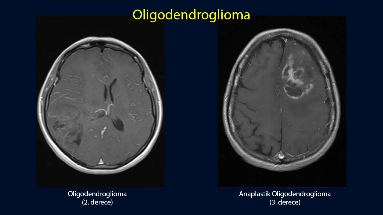 Grade 2 oligodendroglioma on the left, grade 3 on the right. The grade 2 tumor appears more localized and less involved with surrounding tissue; in the grade 3 tumor, the boundaries are less distinct and spread into surrounding brain tissue is more evident. However, MRI alone does not determine the grade: the definitive grade is established by pathological examination of tissue obtained through surgery or biopsy. Contrast enhancement is suggestive of grade 3 but is not required; nonenhancing grade 3 tumors also occur.
2. Tissue Sample
The diagnosis can be established only with tissue. Tissue can be obtained in two ways: during surgical removal of the tumor (the preferred approach, because it provides both diagnosis and treatment), or, if the tumor is in an inoperable location, through stereotactic biopsy.
3. Molecular Testing: A Requirement for Diagnosis
Microscopic examination by the pathologist is not sufficient on its own. The following tests must be performed:
- IDH1 R132H immunohistochemistry —a quick, inexpensive first step
- If staining is negative, IDH1/IDH2 sequencing —to detect rare mutations
- 1p/19q codeletion analysis —using a method capable of demonstrating whole-arm loss (FISH or molecular methods)
- ATRX and p53 staining—to distinguish it from astrocytoma (ATRX is retained in oligodendroglioma)
- MGMT promoter methylation —if chemotherapy is planned
- TERT promoter mutation —supportive
- Ki-67 proliferation index —helps with grading
For more information about these tests and how they are performed, read our article on genetic testing in cancer.
Treatment
Oligodendroglioma treatment has three pillars: surgery, radiotherapy and chemotherapy. Since 2024, a fourth option has been added: the IDH inhibitor vorasidenib. Their sequence and timing depend on the tumor grade and the patient's risk group.
1. Surgery
The first and most important treatment step. The aim is to remove as much tumor as possible while preserving brain function. The amount removed is strongly associated with survival: compared with biopsy alone, complete removal reduces the risk of death by approximately one third.
For tumors adjacent to speech or movement areas, awake craniotomy may be performed: the patient is awakened during part of the operation, and speech and movement are tested to define the safe limits of removal.
2. After Surgery: Immediate Treatment or Surveillance?
This is the most difficult decision in grade 2 oligodendroglioma. Radiotherapy and chemotherapy delay the disease, but they have long-term cognitive side effects that a young patient may carry for decades. Patients are therefore divided into risk groups.
| Risk group | Definition | Approach |
|---|---|---|
| Low risk | Under 40 and complete tumor removal | Close MRI surveillance is a reasonable option |
| High risk | 40 years of age or older (regardless of the extent of removal) or incomplete removal / biopsy alone | Postoperative treatment is recommended |
This distinction comes from the RTOG 9802 trial. In 251 patients with high-risk grade 2 glioma, adding PCV chemotherapy to radiotherapy increased median survival from 7.8 to 13.3 years; 10-year survival increased from 40% to 60%. The benefit was even more pronounced in the oligodendroglioma subgroup.
3. Radiotherapy
| Dose | Fractions | |
|---|---|---|
| Grade 2 | 50.4 Gy (alternative: 54 Gy) | 28 sessions (alternative: 30) |
| Grade 3 | 59.4 Gy | 33 sessions |
According to the 2024 guideline from the European radiation oncology and neuro-oncology societies (ESTRO-EANO), in grade 3, 60 Gy should not be exceeded, and low doses such as 45 Gy are not recommended for grade 2. For general information on how radiotherapy is delivered, see our related article.
4. Chemotherapy: PCV
PCV combines three drugs and is the only chemotherapy regimen proven in randomized trials to prolong survival in oligodendroglioma.
| Drug | Dose | Day | Route |
|---|---|---|---|
| Procarbazine | 60 mg/m² | Days 8-21 | Oral |
| CCNU (lomustine) | 110 mg/m² | Day 1 | Oral |
| Vincristine | 1.4 mg/m² | Days 8 and 29 | Intravenous |
Each cycle lasts 8 weeks, with a total of 6 cycles planned.
PCV or Temozolomide?
In clinical practice, temozolomide is often used instead of PCV because it is easier to tolerate. However, honesty is needed: this choice rests on convenience rather than evidence.
- In patients with 1p/19q codeletion, PCV is the only regimen shown in randomized trials to prolong survival.
- Temozolomide has never been shown to be equivalent to PCV.
- CODEL originally included a temozolomide-only arm. Because outcomes in that arm were substantially worse than in the radiotherapy-containing arms, it was closed.
- The redesigned CODEL compares radiotherapy + PCV with radiotherapy + temozolomide, and its results have not yet been announced.
Vorasidenib: A New Step in Treatment
For years, after surgery for oligodendroglioma, the only options were surveillance or radiotherapy + chemotherapy. The first drug to bridge the gap—"treatment is needed, but I do not yet want to give radiotherapy"—is vorasidenib (brand name Voranigo).
Vorasidenib blocks mutant IDH1 and IDH2 enzymes and is able to enter the brain. Taken orally, it slows tumor growth by stopping abnormal 2-HG production.
The INDIGO Trial
A placebo-controlled, double-blind phase 3 trial involving 331 patients. These were patients who had undergone surgery alone without prior radiotherapy or chemotherapy, for grade 2 IDH-mutant glioma; 172 had oligodendroglioma.
The updated analysis published in 2025 (median follow-up 20.1 months) added two striking figures:
- Tumor growth rate: in the vorasidenib arm, -1.3% (meaning the tumor shrank), compared with the placebo arm, +14.4%.
- Seizure frequency: per person-year, 18.2 versus 51.2 —vorasidenib substantially reduced seizures.
There was no difference between the two arms in quality of life or cognitive testing.
Approvals and Use
| Item | Information |
|---|---|
| FDA approval | 6 August 2024 —after surgery, in patients aged 12 and older with IDH1/IDH2-mutant grade 2 astrocytoma or oligodendroglioma |
| European Union approval | 22 September 2025 —grade 2 IDH-mutant diffuse glioma in adults and adolescents |
| Dose | Adults: 40 mg orally once daily. In patients older than 12 who weigh under 40 kg, 20 mg. |
| Guideline position | ASCO and the Society for Neuro-Oncology's rapid update dated 30 April 2025: in grade 2 oligodendroglioma, following surgery, when radiotherapy and chemotherapy can be deferred, it may be offered (high-quality evidence, conditional recommendation). |
| Use in Grade 3 | Not approved. INDIGO included only patients with grade 2 disease. |
Survival: Current Figures
This section shows the greatest difference between older sources and current data. Most figures circulating online come from the era before molecular diagnosis and do not represent today's patients.
| Group | Current median survival | Old/incorrect figure |
|---|---|---|
| Grade 2 | 15-20 years (18.4 years in a modern series; 10-year survival 76%). Even in high-risk patients, 13.3 years with RT+PCV. | "10-12 years" |
| Grade 3 | 13-14 years (RT + PCV, 1p/19q-codeleted) | "3.5 years" |
Other Factors Affecting Outcomes
| Factor | Effect |
|---|---|
| Removal of nearly all the tumor | Favorable —approximately one-third reduction in the risk of death |
| Younger age | Favorable |
| A seizure at diagnosis | Favorable (effect of early diagnosis) |
| Contrast enhancement on MRI | Unfavorable |
| High Ki-67 index (above 20%) | Unfavorable |
| CDKN2A/B homozygous loss | Unfavorable (approximately 2.4-fold risk) |
| Absent MGMT methylation | In patients receiving chemotherapy, unfavorable |
| Good performance status | Favorable |
Seizure Management
- Preventive antiseizure medication is not started in patients who have never had a seizure. Neuro-oncology societies have issued a clear recommendation (level A): patients with brain tumors who have never had a seizure should not be started on medication to reduce seizure risk.
- If preventive medication was used around surgery, it is usually tapered and stopped at the end of the first week.
- For patients who have had a seizure, the preferred drug is generally levetiracetam: it has few interactions with chemotherapy because it does not affect liver enzymes, and its side effect profile is better than that of older drugs.
- Older enzyme-inducing antiseizure drugs (phenytoin, carbamazepine) can lower chemotherapy drug levels in the blood and are avoided when possible.
- The reduction in seizure frequency by more than half with vorasidenib shows that tumor control and seizure control are linked.
- Driving licenses: for patients who have had seizures, driving requires a specified seizure-free period under regulations also in force in Turkey. This is one of the most overlooked practical aspects of living with the disease and should be discussed with your physician.
Follow-Up
Oligodendroglioma requires follow-up over many years. Monitoring is performed with MRI.
| Situation | MRI frequency |
|---|---|
| Grade 2 | For the first 5 years, every 3-6 months, then at least every 6-12 months |
| Grade 3 | For the first 3 years, every 2-4 months, then every 3-6 months indefinitely |
Frequently Asked Questions
- Is Oligodendroglioma Benign or Malignant?
- It is a malignant tumor, although "malignant" can be misleading here. Among brain tumors, it has one of the most favorable prognoses, and a substantial proportion of patients live for decades. It grows slowly, but because its boundaries are indistinct, complete eradication is usually impossible; it is therefore not considered benign either.
- Can I Be Completely Cured?
- A complete cure is generally not expected with current treatments; the goal is to control the disease for many years. However, in twenty-year data, approximately one third of patients are still alive, and most of them lead normal lives. Oligodendroglioma should be thought of as a "chronic disease."
- Can I Continue Working, Driving and Traveling?
- Most patients continue their normal lives outside treatment periods. Driving depends on seizure status and is subject to legal regulations. Work capacity varies with tumor location and cognitive effects; tasks requiring attention and planning can become difficult in patients with frontal lobe involvement.
- Can I Pass It On to My Children?
- No. IDH mutation and 1p/19q loss are acquired changes arising in brain cells during life; they are absent from reproductive cells and are not inherited.
- Is Surveillance Without Any Treatment Acceptable After Surgery?
- Yes. In low-risk grade 2 patients (under 40, with nearly all the tumor removed), close MRI surveillance is a legitimate option. The aim is to delay the long-term cognitive side effects of radiotherapy as much as possible. The decision should be reviewed at every MRI; this is active surveillance, rather than "doing nothing."
- Could I Receive Vorasidenib?
- The approval covers grade 2 IDH-mutant glioma in patients who have undergone surgery but not yet radiotherapy/chemotherapy. It is not approved for grade 3. Discuss access in Turkey with your treatment team.
- Will Radiotherapy Affect My Memory?
- Brain radiotherapy can cause some decline in attention, processing speed and memory, particularly over the years. This risk underlies the discussion about delaying treatment. Modern techniques (intensity-modulated radiotherapy and hippocampal-sparing planning) aim to reduce this effect. Its extent varies greatly between individuals, and most patients can continue their daily lives.
- Can Diet or Supplements Affect the Tumor?
- No diet, herbal product or supplement has been shown to shrink oligodendroglioma or stop its progression. Some herbal products affect liver enzymes and can change blood levels of chemotherapy and vorasidenib; tell your physician about every product you use.
- What Happens If It Recurs?
- Options include repeat surgery, reirradiation and chemotherapy regimens not used previously (PCV or temozolomide, whichever has not been given). Decisions are made according to the location of recurrence, prior treatments, elapsed time and the patient's general condition, on an individual basis.
Glossary
- IDH mutation
- A change in the isocitrate dehydrogenase enzyme. It is an early event in gliomas and the strongest predictor of a favorable outcome.
- 1p/19q codeletion
- Simultaneous complete loss of the short arm of chromosome 1 and the long arm of chromosome 19. The defining feature of oligodendroglioma.
- Codeletion
- Simultaneous loss of two genetic regions.
- Ki-67
- A stain showing what proportion of cells are dividing; it reflects tumor growth activity.
- MGMT methylation
- Silencing of a DNA repair gene. Alkylating chemotherapy is more effective when it is silenced.
- Gross total resection (GTR)
- Removal of all visible tumor on imaging.
- Pseudoprogression
- A temporary reaction after radiotherapy that makes the tumor appear larger on MRI without true progression.
- Gy (Gray)
- A unit of radiation dose.
- Median survival
- The period exceeded by half of patients; it is not an individual's allotted lifespan.
- Louis DN, et al. The 2021 WHO Classification of Tumors of the Central Nervous System: a summary. Neuro-Oncology 2021;23(8):1231-51.
- cIMPACT-NOW Update 12: grading and risk stratification of IDH-mutant diffuse gliomas. Neuro-Oncology 2026 (advance online publication).
- Price M, et al. CBTRUS Statistical Report: Primary Brain and Other Central Nervous System Tumors Diagnosed in the United States in 2018-2022. Neuro-Oncology 2025;27(Suppl 4):iv1-iv66.
- Lassman AB, et al. Joint Final Report of EORTC 26951 and RTOG 9402. J Clin Oncol 2022;40(23):2539-45.
- Cairncross G, et al. Phase III trial of chemoradiotherapy for anaplastic oligodendroglioma: long-term results of RTOG 9402. J Clin Oncol 2013;31(3):337-43.
- van den Bent MJ, et al. Adjuvant procarbazine, lomustine, and vincristine chemotherapy in newly diagnosed anaplastic oligodendroglioma: long-term follow-up of EORTC 26951. J Clin Oncol 2013;31(3):344-50.
- Buckner JC, et al. Radiation plus Procarbazine, CCNU, and Vincristine in Low-Grade Glioma. N Engl J Med 2016;374:1344-55 (RTOG 9802).
- Mellinghoff IK, et al. Vorasidenib in IDH1- or IDH2-Mutant Low-Grade Glioma. N Engl J Med 2023;389(7):589-601 (INDIGO).
- Cloughesy TF, et al. Vorasidenib in IDH-mutant grade 2 glioma: secondary and exploratory endpoints of the INDIGO trial. Lancet Oncol 2025;26(12):1665-75.
- FDA: vorasidenib approval, 6 August 2024; VORANIGO prescribing information. European Commission approval, 22 September 2025.
- Blakeley J, et al. ASCO-SNO Rapid Recommendation Update: vorasidenib. Neuro-Oncology 2025;27(6):1412-18.
- Baumert BG, et al. ESTRO-EANO guideline on target delineation and radiotherapy for IDH-mutant WHO CNS grade 2 and 3 diffuse glioma. Radiother Oncol 2024.
- Weller M, et al. EANO guidelines on the diagnosis and treatment of diffuse gliomas of adulthood. Nat Rev Clin Oncol 2021;18:170-86.
- Walbert T, et al. SNO and EANO practice guideline update: anticonvulsant prophylaxis in patients with newly diagnosed brain tumors. Neuro-Oncology 2021;23(11):1835-44.
- Mortazavi A, et al. IDH-mutated gliomas promote epileptogenesis through d-2-hydroxyglutarate-dependent mTOR hyperactivation. Neuro-Oncology 2022;24(9):1423-35.
- Wen PY, et al. RANO 2.0: Update to the Response Assessment in Neuro-Oncology Criteria. J Clin Oncol 2023;41(33):5187-99.
- Kinslow CJ, et al. Extent of resection and survival for oligodendroglioma: a US population-based study. J Neurooncol 2019;144(3):591-601.
- Keogh RJ, et al. Real-world tolerability of PCV chemotherapy. BMC Cancer 2021;21:140.
- Jaeckle KA, et al. CODEL: phase III study of RT, RT+TMZ, or TMZ for newly diagnosed 1p/19q codeleted oligodendroglioma. Neuro-Oncology 2021;23(3):457-67.
- Nakasu S, et al. Prognostic significance of homozygous deletion of CDKN2A/B in oligodendroglioma: a meta-analysis. Neurol Med Chir (Tokyo) 2024;64(12):442-50.
- Byeon S, et al. Long-term outcomes of grade 2 oligodendroglioma. Discover Oncology 2024;15:268.
Sources
https://www.cancer.gov/rare-brain-spine-tumor/tumors/oligodendroglioma
2. Tork CA, et al. (2023). Oligodendroglioma.
https://www.ncbi.nlm.nih.gov/books/NBK559184
3. Zerdan MB, et al. (2021). Oligodendroglioma: A review of management and pathways.
https://www.ncbi.nlm.nih.gov/pmc/articles/PMC8523914/
Was this article helpful?
Your feedback helps us improve our content.




Reader comments
Share your comment
Your name and comment are published only after administrator approval. Your email address is never shown to visitors.
Be the first to comment.