
Anal Cancer Symptoms, Causes and Treatment

In this article
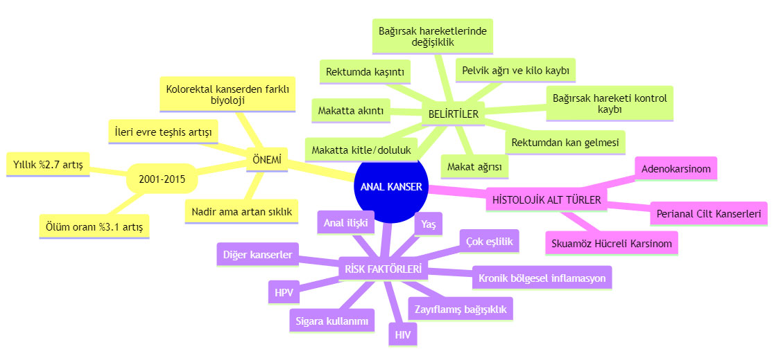
Although anal cancer is rare, both its incidence and mortality are increasing. United States data between 2001 and 2015 show annual increases of 2,7 percent in incidence and 3,1 percent in mortality. Anal cancer is thus among the fastest-rising cancers in both measures in the United States. The stage distribution also changed during this period: advanced-stage diagnoses tripled. Proper management is therefore extremely important.
Most anal cancers are squamous cell carcinomas, and more than 90% of squamous cell anal cancers are caused by human papillomavirus (HPV). A United States study found that more than 75% of adults did not know HPV could cause cancer. Yet HPV infection is a leading preventable cancer cause.
What Is Anal Cancer?
Anal cancers develop in the 2,5-3,5 cm anus at the end of the digestive tract. Anal cancer differs from colorectal cancer and is much rarer, but new cases are increasing, particularly in Western populations. Various risk factors increase the likelihood, although nearly 90% are caused by human papillomavirus (HPV). Most occur after 60 and are more common in women than men.
The infographic below visually presents anal cancer’s importance, symptoms, risk factors and types.
What Are Anal Cancer Symptoms?
Anal cancer may not always cause symptoms. Generally, of 10 cases, 2 are asymptomatic. Possible symptoms include:
- Rectal bleeding: One of the most common symptoms. Bleeding may be clearly visible from the anus or noticed as blood traces on stool. Dark or hard stools may indicate bleeding.
- Itching in or around the rectum: Anal itching has many causes. Anal cancer is generally asymptomatic, meaning it is usually not associated with anal itching.
- A lump or fullness in the anus: As anal cancer grows, a lump or fullness may develop, more commonly in advanced stages.
- Anal pain: Anal cancer may cause pain, but it can resemble pain from other conditions.
- Changes in bowel movements and narrowing stools: Anal cancer can alter bowel movements and narrow stools, but these symptoms resemble other bowel problems.
- Anal discharge: Anal cancer may cause discharge, but it can resemble discharge from other conditions.
- Loss of bowel control: Anal cancer may cause difficulty or loss of bowel control, but this also resembles other bowel problems.
- Other symptoms such as pelvic pain and weight loss: These may occur as anal cancer progresses.
Sometimes specialists identify swollen lymph nodes in the groin or around the anus during examination.
Tumors in the anus may be associated with inflammatory responses to infections such as human papillomavirus (HPV) 16 and 18. People with anal HPV have a 40-fold higher anal cancer risk. Men with genital viral infections also have increased risk.
Anal cancer usually causes no symptoms until advanced. Its symptoms are difficult to distinguish from hemorrhoids, so people experiencing itching should consult a specialist.
Disorders That Can Occur in the Anal Region are illustrated below.

What Are Anal Cancer Causes / Risk Factors?
Many risk factors are linked to anal cancer. The main ones are:
- Human Papillomavirus (HPV): Some HPV types are closely related. Approximately 79% of anal cancer patients have HPV 16 or 18, and 8% other HPV types.
- Multiple Sexual Partners: Having multiple sexual partners increases anal cancer and HPV risk.
- Anal Intercourse: Anal intercourse increases risk in men and women. HIV-positive gay men have a 90-fold higher risk than the general population.
- Other Cancers: Women with vaginal or cervical cancer and men with penile cancer have increased anal cancer risk, also linked to HPV infection.
- Age: Like most cancers, anal cancer risk rises with age.
- Weakened Immune System: People with HIV or AIDS and those taking immunosuppressive drugs after organ transplantation have increased risk.
- Smoking: Smokers have much higher risks of anal and other cancers than nonsmokers.
- Chronic Local Inflammation: Benign anal conditions such as hemorrhoids or fistulas may increase risk through the inflammation they cause.
What Are the Types of Anal Cancer?
- Squamous Cell Carcinoma: Of anal cancers, 90% are squamous cell carcinoma (SCC), starting in squamous cells lining most of the anal canal and lower anus.
- Adenocarcinoma: This starts in cells forming the upper anus or mucus-producing glands beneath the anal lining.
- Perianal Skin Cancers: Cancers in skin around the anus, no more than 5 cm away, are classified as perianal skin cancers.
How Is Anal Cancer Diagnosed?
Diagnosis involves digital rectal examination, anoscopy and biopsy of an identified mass. Anoscopy uses an anoscope (a lighted tube) to examine the anal canal and rectum. Biopsy takes tumor tissue samples for microscopic examination for cancer. It both confirms diagnosis and determines characteristics such as histological subtype.
When an anal tumor is found, its anatomical location must be defined. The origin of tumors overlapping the anorectal junction can be difficult to identify. Generally, tumors more than 2 cm proximal to the dentate line or proximal to the anorectal ring are classified as rectal cancer; those 2 cm or less proximal to the dentate line as anal cancer. Skin-origin cancers at or distal to the squamous mucocutaneous junction and skin cancers no more than 5 cm from the anus are classified as perianal skin cancers.
Once anal cancer is confirmed, advanced imaging determines its stage and whether it has spread (metastasized), for example magnetic resonance imaging and PET tomography .
How Is Anal Cancer Staged?
As with other cancers, treatment is planned by stage. Staging assesses tumor size, invasion of adjacent structures, lymph node metastases and distant metastases. Evaluations include physical examination and biopsy of the primary tumor, gynecological and groin examinations, PET, chest CT, and abdominal/pelvic CT or MRI.
Below, TNM staging of anal cancer is shown:

The final TNM stage classification is:
- Stage 1: Tumor diameter is 2 cm or less.
- Stage 2: Tumor diameter exceeds 2 cm.
- Stage 3: The tumor has spread to adjacent organs such as the vagina, urethra or bladder, or lymph nodes near the pelvis/rectum.
- Stage 4: Cancer has spread (metastasized) to distant sites such as lungs, liver or brain.

Anal Cancer Treatment
What factors determine treatment?
Treatment planning first establishes tumor location, histological subtype and stage. Once tumor features are defined, patient factors affecting decisions are assessed: age, performance status, comorbidities and HIV positivity.
Patients with HIV
HIV-associated anal SCC is treated the same as in uninfected patients, with similar local control, response and survival. Low CD4 counts do not increase treatment-related toxicity risk. However, patients with active HIV/AIDS complications or a history of complications, such as opportunistic infections or other HIV-related cancers, may not tolerate full doses or mitomycin, a standard chemotherapy agent. Dose adjustments or mitomycin-free treatment may be needed.
Preclinical data suggest similar tumor microenvironments in HIV-positive and HIV-negative patients; immunotherapy decisions in metastatic anal SCC should therefore be made regardless of HIV status.
Older Patients or Those with Extensive Comorbidities
Standard treatment remains concurrent chemotherapy and radiotherapy (chemoradiotherapy - CRT), as in younger patients without comorbidities. Although trials are limited, reducing concurrent mitomycin and fluorouracil (FU) doses may be considered. In older or seriously comorbid patients with T1N0 tumors, FU alone with radiotherapy, omitting mitomycin, may be considered.
How Are Non-Squamous Cancers in the Anal Region Treated?
Perianal Skin Cancer
For well-differentiated T1N0 perianal squamous cell skin cancers (less than 2 cm, no spread), clearly separate from the anal canal, wide local excision alone is sufficient if negative margins can be achieved while preserving sphincter muscles—meaning complete removal. Postoperative radiotherapy is an option with high-risk histological features such as poor differentiation or perineural invasion.
If the tumor exceeds 2 cm, surgery threatens sphincter function, lymph nodes are involved or anatomical location cannot be clearly separated from the anal canal, FU- and mitomycin-based chemoradiotherapy is recommended, reserving surgery for persistent or recurrent disease.
Anal Adenocarcinoma
Localized anal canal adenocarcinoma should be treated like localized rectal adenocarcinoma, with surgery (abdominoperineal resection - APR) and FU-based chemoradiotherapy before or after surgery, rather than FU plus mitomycin as for localized anal SCC.
Rectal Squamous Cell Carcinoma
These cancers are regarded as anal squamous cancers extending into the distal rectum and treated like anal SCC, with FU- and mitomycin-based chemoradiotherapy.
How Is Anal Squamous Cell Cancer Treated?
Treatment of Localized Disease
Concurrent chemoradiotherapy is standard for localized anal SCC, even stage 1. Although no randomized trial directly compares it with surgery, CRT preserves the sphincter and cures 70-85% of patients. Local excision may be an option in carefully selected superficial invasive tumors with basement membrane invasion of 3 mm or less, maximum horizontal spread of 7 mm or less and size below 1 cm. However, studies have not compared local excision with radiotherapy or chemoradiotherapy. If chosen, close follow-up and immediate CRT on recurrence are essential.
Initial CRT preferably combines radiotherapy with FU and mitomycin. NCCN recommends the RTOG 98-11 protocol: days 1-4 and 29-32, infusional FU 1000 mg/m2 , with days 1 and 29 mitomycin 10 mg/m2 (maximum 20 mg per dose). ESMO recommends the ACT II protocol: days 1-4 and 29-32, infusional FU 1000 mg/m2 , with only day 1 single-dose mitomycin 12 mg/m2 (maximum 20 mg). Both protocols are applicable. NCCN and ESMO also recommend FU+ cisplatin or mitomycin+capecitabine as alternative concurrent chemotherapy regimens with radiotherapy.
Treatment of Locally Advanced Disease
For locally advanced anal SCC (T3/4 or N1b/c), standard treatment similarly combines radiotherapy with FU and mitomycin. Only the total radiation dose differs, with 10-14 Gy more recommended (45 versus 55-59). Standard chemoradiotherapy achieves cure in 50-60%.
Treatment of Metastatic Disease
Systemic therapy is standard for metastatic anal SCC. A small subgroup with isolated liver metastases may benefit from resection, but selection criteria have not been defined.
Isolated para-aortic lymph node involvement is rare and considered M1 (stage IV) disease. Optimal treatment is undefined, but CRT including these nodes in the radiation field is reasonable. These patients remain at high risk for additional metastases.
Systemic Chemotherapy
Both ESMO and NCCN recommend carboplatin+paclitaxel as the preferred first-line regimen for metastatic anal SCC. Cisplatin+FU and modified DCF (docetaxel, cisplatin, FU) are alternatives.
InterAACT randomized 91 previously untreated metastatic anal SCC patients to carboplatin (AUC=5, every 28 days)+weekly paclitaxel (80 mg/m2, days 1, 8 and 15, every 28 days), or cisplatin (60 mg/m2, every 21 days)+infusional FU (1000 mg/m2, days 1-4, every 21 days), forming 2 groups. Although response rates, the primary endpoint, were similar (59% versus 57%), overall survival (20 versus 12,3 months) and toxicity (serious side effects 36% versus 62%) were significantly better with carboplatin+paclitaxel.
Besides carboplatin every 28 days on 1 day with weekly paclitaxel, a regimen giving both drugs every 3 weeks on 1 day is widely used. Differences in treatment toxicity between these protocols are unknown, but both are reasonable.
Immunotherapy
Single-agent immunotherapy targeting the programmed cell death receptor-1 (PD-1) pathway is recommended for metastatic anal SCC progressing after first-line treatment. Optimal criteria for selecting those most likely to benefit remain undefined; testing PD-L1 overexpression is therefore not currently recommended. Trials combining these agents with cytotoxic chemotherapy first-line are ongoing, but no clear data are yet available.
Promising results in metastatic SCC have been reported with PD-1 blockade using nivolumab, pembrolizumab and retifanlimab. Without assessment of PD-1 expression, a small phase II study of 37 chemotherapy-resistant metastatic anal SCC patients administered a median 6 cycles (3-10 cycles) of 3 mg/kg intravenous (IV) nivolumab every two weeks. Results: 9 patients (24%) had objective responses, 2 (5%) complete responses and 17 (46%) stable disease as best response. Median progression-free survival was 4,1 months and overall survival 11,5 months. In PD-L1-positive anal SCC, 24 patients were assessed in KEYNOTE-028: pembrolizumab every 2 weeks at 10 mg/kg IV for up to two years produced partial responses in 4 (17%) and stable disease as best response in 10 (42%). After first-line progression, metastatic or inoperable anal SCC in 112 patients was evaluated in phase II KEYNOTE-158. Patients received treatment for 2 years or until progression, every 3 weeks with 200 mg IV pembrolizumab. At median follow-up of 34,7 months, 12 patients had objective responses (11%), and 6 (5%) complete responses. However, median progression-free survival was only 2,1 months and overall survival 11,9 months. After platinum-based first-line progression, locally advanced or metastatic anal SCC in 94 patients was assessed in phase 2 POD1UM-202: treatment every 4 weeks with 500 mg IV retifanlimab yielded 14% objective response (1 complete response), 13% partial response and 35% stable disease; disease control was 49%. Median response duration was 9,5 months, progression-free survival 2,3 months and overall survival 10,1 months.
How Is Anal Cancer Followed Up?
No prospective study guides post-treatment anal SCC follow-up. Most recurrences occur within the first three years, requiring closer monitoring. NCCN recommends reassessment 8-12 weeks after CRT by digital rectal examination (DRE), classifying response as complete, persistent or progressive disease. Complete responders should undergo DRE, anoscopy and inguinal lymph node examination every 3-6 months for five years. Annual contrast-enhanced chest, abdominal and pelvic CT or MRI is also recommended for 3 years.
Evidence is insufficient to recommend fluorodeoxyglucose positron emission tomography (FDG-PET/CT) for assessing response or following treated anal cancer.
What If Persistent / Recurrent Disease Is Found During Follow-Up?
Risk factors include primary tumor size >5 cm, lymph node involvement, poor adherence, unnecessary chemotherapy dose reductions and treatment delays.
Persistent or locally recurrent disease after CRT can be successfully treated by surgery (abdominoperineal resection - APR), but long-term control is achieved in only 24-40% of locally recurrent anal SCC.
APR is also preferred when disease persists after CRT. Based on ACT II, patients with suspected persistence at the first examination 8-12 weeks after treatment can be observed for up to six months after radiotherapy and chemotherapy if no progression occurs. Progression at any time, or suspected persistent disease six months or more after CRT, requires biopsy; confirmed persistence or recurrence warrants salvage APR.
The effectiveness of nonsurgical salvage treatment to preserve the sphincter in persistent disease is unknown. A small study of 22 patients reported 4 long-term disease-free survivors after salvage CRT with FU and cisplatin, but evidence is insufficient for firm conclusions. Continuing sphincter-preserving treatment for persistent/recurrent disease is therefore a difficult decision requiring multidisciplinary assessment.
Sources
2. Rao S, Sclafani F, Eng C et al. International Rare Cancers Initiative Multicenter Randomized Phase II Trial of Cisplatin and Fluorouracil Versus Carboplatin and Paclitaxel in Advanced Anal Cancer: InterAAct. J Clin Oncol. 2020; 38:2510.
3. Ott PA, Piha-Paul SA, Munster P, et al. Safety and antitumor activity of the anti-PD-1 antibody pembrolizumab in patients with recurrent carcinoma of the anal canal. Ann Oncol. 2017; 28:1036.
4. Marabelle A, Cassier PA, Fakih M, et al. Pembrolizumab for previously treated advanced anal squamous cell carcinoma: results from the non-randomised, multicohort, multicentre, phase 2 KEYNOTE-158 study. Lancet Gastroenterol Hepatol. 2022; 7:446.
5. Welton ML, Steele SR, Goodman KA, et al. Anus. In: AJCC Cancer Staging Manual, 8th ed, Amin MB (Ed), AJCC, Chicago 2017. p.275.
6. Lam AK, Goldblum JR. Tumours of the anal canal: Introduction. In: WHO Classification of Tumours: Digestive System Tumours, 5th ed, WHO Classification of Tumours Editorial Board (Ed), International Agency for Research on Cancer, Lyon 2019.
7. NCCN Clinical Practice Guidelines in Oncology, Anal Carcinoma, Version 3.2023. Available at:
https://www.nccn.org/professionals/physician_gls/pdf/anal.pdf (Accessed Oct 31, 2023)
8. Rao S, Guren MG, Khan K, et al. Anal cancer: ESMO Clinical Practice Guidelines for diagnosis, treatment and follow-up. 2021; 32: p1087-1100.
9. Rao S, Anandappa G, Capdevila J, et al. A phase II study of retifanlimab (INCMGA00012) in patients with squamous carcinoma of the anal canal who have progressed following platinum-based chemotherapy (POD1UM-202). ESMO Open. 2022; 7:100529.
Was this article helpful?
Your feedback helps us improve our content.




Reader comments
Share your comment
Your name and comment are published only after administrator approval. Your email address is never shown to visitors.
Be the first to comment.