
Lung Cancer Staging Has Changed! Let Us Review Its Symptoms and Treatment

In this article
There are two main lung cancer types: non-small-cell and small-cell lung cancer. Each has its own staging system. Staging establishes the extent of spread, helps understand the disease’s phase and enables doctors to develop the best possible treatment plan.
This article discusses:
- Lung cancer stages,
- Symptoms and screening for early diagnosis,
- Diagnosis and treatment
Determining lung cancer stages
Both main lung cancer types have their own staging systems.

Most lung cancers (approximately %80) are non-small-cell lung cancer (NSCLC).
Non-small-cell lung cancer – NSCLC
Non-small-cell lung cancer (NSCLC) accounts for approximately 80 percent of cases and has three main subtypes:
- Adenocarcinoma: Accounts for 40 percent of lung cancers. Usually in peripheral/outer lung regions, it tends to grow more slowly than the other two subtypes and is therefore more likely to be detected before metastasis.
- Squamous cell carcinoma: Accounts for approximately 25-30 percent. It arises from cells lining the airways, usually the bronchi, and tends to be centrally located.
- Large-cell carcinoma: Accounts for 10-15 percent, occurs anywhere in the lung and tends to grow and spread faster than other subtypes.

NSCLC is most commonly classified using TNM as its staging system:
T – tumor size and location. Tumor size has gained importance because it directly affects disease course and survival. Tumors smaller than 1 cm are classified T1a (previously those smaller than 2 cm were T1a).
Not all small tumors are treated alike; many early-stage options exist, for example SBRT, surgery and cryoablation. Molecular profiles and genetic characteristics must also be assessed.
N – number of involved lymph nodes. The N category is unchanged in the new system.
M – metastasis or cancer spread. This system better shows that not all metastatic NSCLCs are alike and that the number of distant sites matters. A single metastasis outside the chest is M1b, multiple metastases M1c. With good treatment planning, prognosis is better in patients with a single metastasis, also called “oligometastasis.”
Combining T, N and M scores classifies NSCLC into four stages:
- Stage 1: the tumor is in one lung with no spread to lymph nodes or distant organs.
- Stage 2: cancer has spread to lymph nodes within the lung but not distant organs.
- Stage 3: cancer involves central chest (mediastinal) nodes but not distant organs.
- In stage 3a, cancer has not spread to the opposite lung.
- In stage 3b, it has spread to lymph nodes in the opposite lung and toward the head and neck up to the collarbone.
- Stage 4: cancer has spread through the body (metastatic stage).
Small-cell lung cancer
Small-cell lung cancer (SCLC) accounts for approximately 15 percent of all lung cancers. The most common system divides it into two stages:
- Limited stage: cancer is only on one side of the chest, also called thorax-limited disease.
- Extensive stage: cancer has spread to the opposite lung or elsewhere in the body.
SCLC grows and spreads rapidly, making treatment difficult. With overall 5-year survival of % 5-10, thorax-limited disease requires more comprehensive assessment.
Lung cancer symptoms and screening recommendations for early diagnosis
Symptoms resemble bronchitis or pneumonia, one reason more than % 70 are detected at advanced stages.
However, earlier detection increases the likelihood of successful treatment. Consult a physician if you have:
- Persistent cough or blood in sputum
- Hoarseness, chest pain, shortness of breath or wheezing
- Frequent respiratory infections
- Persistent fatigue or weakness, appetite loss and unexplained weight loss
As cancer progresses, new signs and symptoms may include:
- Headache, dizziness or seizures from brain spread
- Jaundice from liver spread
- Lumps under the skin from lymph node spread
- Back, hip or general bone pain from bone metastases
Although no significant benefit has been shown in low-risk patients, for heavy smokers there is strong evidence that low-dose CT cancer screening can substantially reduce mortality.
Diagnosis and treatment
Suspicious findings prompt computed tomography (CT) or PET-CT scanning. These produce detailed images revealing tumor size, shape and location.
The next step is to determine malignancy and, if cancerous, the lung cancer subtype through biopsy . The tissue sample is examined microscopically and undergoes genetic tests.
Treatment is then planned from the information gathered. It combines methods according to stage, type and tumor genetics.
Traditional treatments are surgery, radiation and chemotherapy:
- Surgery: In early cancer, complete removal of the primary tumor may be possible.
- Radiotherapy: X-rays damage cancer-cell DNA, killing cells and shrinking or destroying tumors.
- Chemotherapy: Drugs injected intravenously interact with cancer-cell DNA to prevent growth and spread.
Radiotherapy and chemotherapy cannot distinguish cancer and healthy cells well, so healthy cells are also partly damaged.
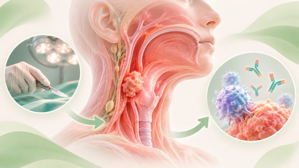
Alongside traditional therapies, new drugs attacking only cancer cells are becoming more accessible. They generally have less severe side effects and significantly prolong survival as new options.
Targeted therapies, smart drugs: These target specific cancer-cell proteins arising from abnormal DNA changes and identifiable through tumor genetic testing.
Immunotherapy: Certain cancer-cell surface proteins interact with proteins on immune cells, like a false friendly hand, preventing immunity from recognizing cancer as harmful. Immunotherapy blocks these interactions, removing the disguise so immunity can attack.

Complete treatment, called cure, is unfortunately not always possible. Supportive methods then relieve symptoms and pain and provide the best possible quality of life. Such treatment is generally called palliative care .
How can I reduce lung cancer risk?
Lung cancer is the leading cause of cancer deaths. Tobacco is unquestionably its greatest risk factor. The best prevention is therefore not smoking or quitting.
Related Topic:
Sources
TNM Classification of Malignant Tumours, 8th ed. Hoboken, New Jersey; Wiley-Blackwell; 2017.
Was this article helpful?
Your feedback helps us improve our content.

Reader comments
Share your comment
Your name and comment are published only after administrator approval. Your email address is never shown to visitors.
Be the first to comment.