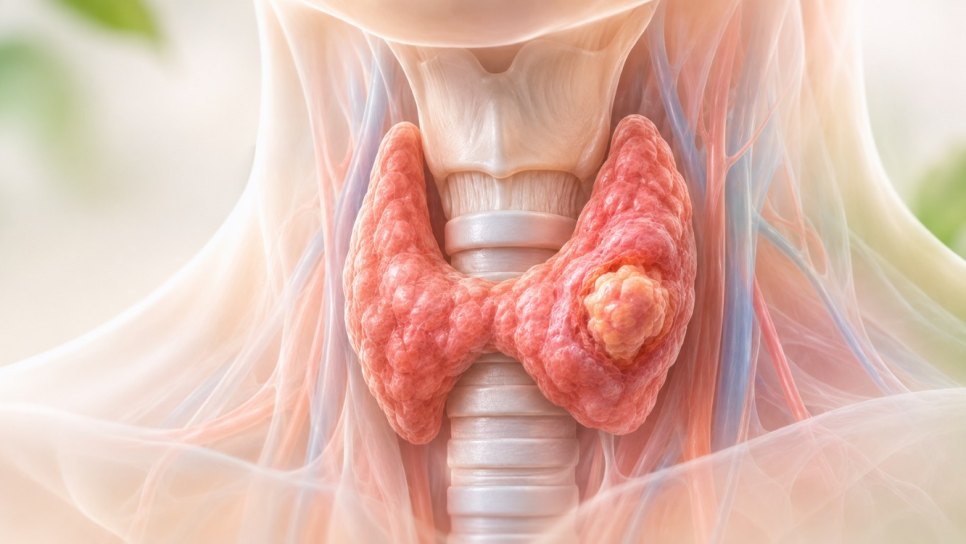
Mitotic Count and Risk Classification in Gastrointestinal Stromal Tumors (GIST): Fletcher Criteria
In this article
If Your Pathology Report Says GIST and “Mitotic Count”: How Is Recurrence Risk Determined?
Gastrointestinal stromal tumor (GIST) is a sarcoma that arises from special cells in the wall of the digestive tract, most often in the stomach and small intestine. One of the most important measures of recurrence risk after surgery is the mitotic count obtained by the pathologist; together with tumor size, location, and tumor rupture it defines the risk group. This article explains how the Fletcher (NIH) and AFIP tables are read, why it matters whether mitoses are counted per “50 high-power fields” or per “5 mm²”, and which evidence underlies the decision on adjuvant (post-operative) imatinib. For background, see our articles on stomach cancer symptoms, stages, and treatment and terms used in pathology reports (in Turkish). Key source: Joensuu et al., Lancet Oncol 2012 (doi:10.1016/S1470-2045(11)70299-6).
- What was found: In high-risk GIST, 3 years of adjuvant imatinib raised 5-year recurrence-free survival from 47.9% to 65.6% compared with 1 year (HR 0.46); about 18 more of every 100 patients remained recurrence-free at 5 years.
- Basis: The risk tables (Fletcher 2002, AFIP 2006) were derived from observational series. For adjuvant imatinib there are three randomized trials (Z9001, SSG XVIII, EORTC 62024); their results are not the same.
- Caution: Mitoses can be counted per “50 HPF” or per “5 mm²”; with wide-field microscopes 50 HPF covers a larger area and can change the risk class. The mitotic count alone does not decide; size, location, and rupture are read together.
- In practice: Ask in which area (5 mm²) mitoses were counted, and ask about location and rupture. The decision on adjuvant imatinib is made by a multidisciplinary team using the risk group and mutation analysis together.
Why is the mitotic count so decisive?
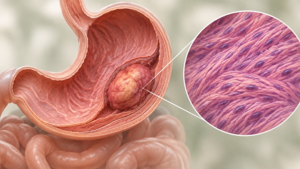
GISTs arise from cells that resemble the interstitial cells of Cajal, which coordinate gut movements; most carry a mutation in the KIT or PDGFRA gene. They are most common in the stomach, then the small intestine, and can also occur in the esophagus, colon, and rectum. A mitosis is the stage of cell division seen under the microscope; the more mitoses are counted in a given area, the faster the tumor is growing.
In practice, risk is determined by four things together: tumor size, mitotic count, location, and tumor rupture. Even at the same size and mitotic count, small-intestinal tumors progress more often than gastric ones, so looking at a single number is misleading. The tumor is usually noticed as a bulge in the wall of the stomach or bowel on endoscopy or imaging (how stomach tumors look on endoscopy); symptoms can include gastrointestinal bleeding and anemia.
The real questions are: In which area and how were mitoses counted, which classification was used, and in which risk group does imatinib after surgery help? Because classifications can give different results, the same patient may receive different “risk” labels in different reports.
No; it has been debated for more than twenty years. What has changed is the standardization of the counting area, the addition of location and rupture, and testing in randomized trials:
How do the Fletcher criteria and the mitotic count determine recurrence risk in GIST?
The Fletcher (NIH) criteria divide GIST into very low, low, intermediate, and high risk groups by tumor size and mitotic count; recurrence becomes more likely as both rise. Current practice counts mitoses per 5 mm² and also considers location and rupture. These are probabilities, not exact predictions; the team decides on adjuvant therapy together with mutation analysis.
How are the Fletcher (NIH) and AFIP tables read?
The table summarizes the risk groups of the Fletcher/NIH 2002 consensus. In the original table the mitotic count is given per 50 high-power fields (HPF); size is the largest diameter of the tumor.
| Risk group | Tumor size | Mitotic count (per 50 HPF) |
|---|---|---|
| Very low | under 2 cm | 5 or fewer* |
| Low | 2–5 cm | 5 or fewer* |
| Intermediate | under 5 cm | 6–10 |
| Intermediate | 5–10 cm | 5 or fewer* |
| High | over 5 cm | more than 5 |
| High | over 10 cm | any count |
| High | any size | more than 10 |
*The original text says “fewer than 5”; AFIP and later systems apply the cut-off as “5 or fewer” / “more than 5”. The AFIP tables apply the same logic separately by location. The figure below shows which four inputs make up the risk group.
Figure 1 · Four inputs that define the risk group
Simplified diagram; not to scale
How much do mitotic count and location change the risk estimate?
AFIP data: same size, different mitoses
In the AFIP tables (Miettinen and Lasota 2006), tumors in the same size group show very different progression rates depending on mitotic count and location. For gastric tumors of 2–5 cm, progression on long follow-up was 1.9% with 5 or fewer mitoses and 16% with more than 5; for the jejunum/ileum at the same size, the figures were 4.3% and 73%. So when mitoses rose, the progression rate increased about 8-fold in gastric tumors and about 17-fold in small-intestinal tumors. “Progression” is the percentage of patients who developed metastasis or tumor-related loss of life.
AFIP 2006: progression on long follow-up in GIST of 2–5 cm
Percent. Lighter bars show 5 or fewer mitoses, darker bars more than 5 mitoses (per 50 HPF).
Retrospective registry series; lower is better. The size of the subgroups cannot be seen from this summary; percentages in small groups can be unstable. The table is reproduced from a secondary publication.
How are mitoses counted? 50 HPF and 5 mm² are not the same
In older sources the mitotic count is given per “50 high-power fields”, and this area was considered roughly equal to 5 mm². Because the field of view of modern microscopes is wider, 50 HPF can correspond to a clearly larger surface than 5 mm². In a survey in Italy, 64.5% of 110 pathologists counted mitoses in 50 HPF and 35.5% in 5 mm². When the same 100 cases were re-evaluated, the risk class changed in 10% of cases at ocular field number 18, 28% at 20, and 41% at 22, when the 50 HPF count was compared with the 5 mm² count. The changes were seen in the very low, low, and intermediate risk groups; the authors note that this could lead to inappropriate adjuvant therapy.
Does the counting area change the risk class?
Italian survey (2022), 100 cases; compared with the 5 mm² count. Values in percent.
50 HPF · field number 18 (≈8 mm²)
50 HPF · field number 20 (≈9.8 mm²)
50 HPF · field number 22 (≈11.9 mm²)
Cases already in the highest class cannot change. The “unchanged” share is calculated to complete 100. A small set of cases (100); the area sizes are approximate values in the source.
Do the classifications agree with each other?
Five classification systems were compared in 558 patients (R0 resection, stomach and small intestine) from the Ulm GIST Registry (Schmieder 2016). All separated high-risk from lower-risk patients well; however, 5-year disease-free survival in the “high risk” group ranged from 56.1% to 74.6% depending on the system. Because the confidence intervals overlapped, the authors reported that no system was significantly superior to the others; the high-risk groups were also heterogeneous within themselves.
Schmieder 2016: 5-year disease-free survival in the “high risk” group
Percent. Each bar is the group of the same 558 patients counted as “high risk” by a different system.
The same patient set, different classifications; because the groups differ in size, the bars do not answer “which is better.” Values are adjusted for tyrosine kinase inhibitor use. Higher is better. Single-center registry.
In the pooled population-based series of Joensuu and colleagues (2,560 patients, 10 series, no adjuvant therapy after surgery), 15-year recurrence-free survival was 59.9% and the number of recurrences declined after the first 10 years. Independent adverse factors were large tumor size, high mitotic count, non-gastric location, tumor rupture, and male sex. For 10-year recurrence-free survival the discrimination (AUC) was 0.79 for NIH, 0.78 for modified NIH, 0.82 for AFIP, and 0.88 for contour maps that model size and mitoses as continuous variables.
Joensuu 2012: AUC for 10-year recurrence-free survival
The closer the AUC is to 1, the better the discrimination (0.5 is chance level). The dark bar is the contour maps.
Pooled 2,560 patients; the reported AUC for the contour maps was 0.88 (95% CI 0.86–0.90). AUC is not a probability; it shows how well a classification separates patients who relapse from those who do not. Higher is better. Validation was also done in an independent cohort of 920 patients.
In conclusion, the modified NIH was judged the best scheme for identifying a single high-risk group to consider for adjuvant therapy, while contour maps were found suitable for individualized recurrence estimates. The mitotic count is a core input of all these models.
How does the risk group translate into treatment?
The mainstay of treatment for localized GIST is complete (R0) removal of the tumor. In patients with a clear risk of recurrence after surgery, imatinib (400 mg/day) is used as adjuvant therapy; the ESMO–EURACAN–GENTURIS guideline recommends continuing it for 3 years and performing mutation analysis before the decision. In low-risk patients, follow-up is usually sufficient.
Figure 2 · The path to the adjuvant imatinib decision
Simplified diagram; the decision is made with the team for each patient
Surgery: why does tumor integrity matter?
The aim of surgery is to remove the tumor with its capsule intact and clear margins. Tumor rupture (tearing or spillage into the abdomen) is an independent factor that markedly raises recurrence risk and enters the risk calculation, so the operative note should state whether rupture occurred. Because lymph node involvement is rare in GIST, routine lymph node dissection is generally not needed. PET-CT is used in some cases to assess spread; the liver is one of the most common sites of metastasis (liver tumors). For advanced disease see stage 4 and metastasis, and for the difference from adenocarcinoma in rectal location see colon and rectal cancer.
Adjuvant imatinib: three randomized trials
The three large trials testing imatinib after surgery cover different patient groups and different comparisons, so placing their results directly side by side would not be correct.
| Trial | Comparison and patients | Finding | Comment |
|---|---|---|---|
| ACOSOG Z9001 (2009) | 1 year of imatinib vs placebo; tumors of at least 3 cm, about 700 patients | 1-year recurrence-free survival 98% vs 83%; HR 0.35 (95% CI 0.22–0.53) | Delayed recurrence on short follow-up; long-term benefit was tested in later trials |
| SSG XVIII (2012; update 2020) | 3 years vs 1 year of imatinib; high risk, 400 patients | 5-year recurrence-free survival 65.6% vs 47.9% (HR 0.46); 10-year overall survival 79.0% vs 65.3% (HR 0.55) | Favors longer treatment in high risk; open-label trial |
| EORTC 62024 (long-term follow-up) | 2 years of imatinib vs observation; intermediate and high risk, 908 patients | 5-year imatinib-failure-free survival 87% vs 83%; HR 0.87 (95.7% CI 0.65–1.15) | No significant difference; a trend in the high-risk subgroup, not significant |
In Z9001, 1 year of imatinib delayed recurrence compared with placebo; in SSG XVIII, 3 years improved both recurrence-free and overall survival compared with 1 year. Absolute difference: 17.7 points in 5-year recurrence-free survival. In EORTC 62024, 2 years of imatinib showed no clear benefit over observation; this trial included intermediate- and high-risk patients together and the duration was shorter than 3 years, so it does not answer the same question as SSG XVIII.
SSG XVIII: 3 years vs 1 year of adjuvant imatinib in high-risk GIST
Percent. Colored bars show the 3-year treatment, gray bars the 1-year treatment (comparison arm).
Arms of the same trial; HR 0.46 for recurrence-free survival. Open-label randomized trial, 400 patients. Higher is better.
In EORTC 62024, after a median follow-up of 9.1 years there was no significant difference between 2 years of imatinib and observation:
EORTC 62024: 2 years of imatinib vs observation in intermediate- and high-risk GIST
Percent, 5-year values. Colored bars show imatinib, gray bars observation only (comparison arm).
Arms of the same trial; 908 patients. HR 0.88 for overall survival (95% CI 0.65–1.21). At 10 years the values were also similar (75% and 74%). Higher is better. Comparison across different trials is indirect.
In the high-risk subgroup (526 patients, local pathology), 10-year imatinib-failure-free survival was 69% vs 61%; this difference was interpreted as a trend but was not statistically significant. No added benefit was seen in intermediate- and low-risk patients. ESMO’s summary of the long-term results also stresses that adjuvant imatinib is recommended in high-risk patients.
Why is mutation analysis as important as risk?
Most GISTs carry a mutation in KIT (most often exon 11) or PDGFRA, and imatinib suppresses the signal these mutations sustain. But not every mutation responds the same way: the PDGFRA D842V mutation is resistant to imatinib, and the benefit of imatinib is limited in SDH-deficient GISTs. Therefore, even when the risk group is high, mutation analysis is recommended before deciding on adjuvant imatinib. For the method, see next-generation sequencing (NGS) and why the location of a mutation matters.
Mutation results cannot be reduced to a single number like the risk tables; detailed results by mutation type are outside the scope of this article. Discuss what the mutation in your report means with the treating team. For access to new drugs see access map of FDA-approved new cancer drugs in Türkiye and, if suitable, clinical trials.
Intermediate risk: the gray zone
The “intermediate” group in the Fletcher table includes tumors under 5 cm with 6–10 mitoses, or tumors of 5–10 cm with a low mitotic count. Decisions are individual in this group: in EORTC 62024 no added benefit was seen in intermediate- and low-risk patients, whereas the modified NIH gathers the candidates for adjuvant therapy into a single high-risk group. Location (small intestine is riskier), rupture, or a mitotic count near the threshold can change the decision; in difficult cases a second opinion is reasonable.
Limits of mitotic counting
Mitoses are counted over an area of 5 mm² in the most active region of the tumor (the “hot spot”); different pathologists, microscopes, and sampling can give different results. Because the whole tumor is not seen on a needle biopsy or gastric biopsy, the mitotic count is only final on the surgical specimen. If imatinib was given before surgery, the mitotic count may be lower, so risk classification in such specimens is interpreted with care. In some small series, proliferation markers such as Ki-67 were also linked to risk groups; however, they are not part of the standard risk calculation.
How should the results be read?
Strengths
- Fletcher, AFIP, and contour maps point in the same direction: recurrence risk rises with size and mitotic count; non-gastric location and rupture raise it further.
- For adjuvant imatinib in high-risk patients there is randomized evidence (Z9001, SSG XVIII) and the difference is clinically large: about 18 points in 5-year recurrence-free survival.
- The contour maps were derived from a pooled series of 2,560 patients and tested in an independent cohort of 920 patients.
- The effect of the mitotic counting area was shown numerically in a separate study (Italy, 110 pathologists, 100 cases).
Points to keep in mind
- Risk tables are observational: The Fletcher and AFIP tables were derived from retrospective series; they give probabilities, not certain predictions.
- Variable counting method: 50 HPF and 5 mm² do not cover the same area; the risk class can change with the counting method.
- Differences between systems: The same patient can fall into different groups in different systems; in the Ulm registry no system was found significantly superior.
- Trials are not comparable: Z9001 (1 year, placebo), SSG XVIII (3 years, 1 year), and EORTC 62024 (2 years, observation) answer different questions; EORTC 62024 showed no significant benefit.
- Mutation distinction: Even when risk is high, no benefit is expected if the tumor carries an imatinib-resistant mutation.
Practical tips for patients and families
If your pathology report says GIST and gives a mitotic count, these steps may help:
- Find the tumor size, location, mitotic count, and the area in which counting was done (5 mm² or 50 HPF) in the report; if missing, ask the pathologist.
- Ask whether the operative note records tumor rupture and whether the surgical margin is clear (R0).
- Ask which risk classification was used (Fletcher/NIH, AFIP, modified NIH); you may see different labels in different reports.
- If adjuvant imatinib is recommended, ask for the mutation analysis result and discuss the duration of treatment and the follow-up plan in writing.
- Seek help early for imatinib side effects (swelling, diarrhea, nausea, fatigue): see our articles on fatigue, diarrhea, and easing side effects with nutrition; the guide to food and drug interactions is also useful.
- Do not skip follow-up; seek care immediately for new abdominal pain, black stools, bleeding, or signs of anemia.
- “If the mitotic count is low, there is no risk.” No. Even with a low count, recurrence risk can be substantial in a large tumor or a non-gastric location; risk is a combination of several inputs.
- “The risk class is single and certain.” No. The same tumor can fall into different groups in different systems and with different counting areas.
- “Imatinib works in every high-risk patient.” Not always. No benefit is expected in PDGFRA D842V and SDH-deficient tumors; the decision is made with mutation analysis.
- “The longer I take it, the better.” 3 years proved superior to 1 year; however, the 2-year EORTC trial did not show superiority over observation. Duration and need are determined with the team according to risk group and mutation.
- We could not confirm that a separate Turkish guideline or reimbursement rule specific to GIST exists; treatment is usually planned in a multidisciplinary tumor board following international guidelines such as ESMO.
- We could not verify, for this article, the marketing authorization of imatinib in Türkiye or the SGK reimbursement conditions for adjuvant GIST use. For general rules see SGK reimbursement conditions for cancer drugs, and for unreimbursed drugs licensed but SGK-unreimbursed cancer drugs (SGK — Turkish Social Security Institution).
- Access to and reimbursement of KIT/PDGFRA mutation analysis and NGS panels may vary by center; we could not verify this, so ask your treatment team.
- Data from Türkiye: in a series of 27 GISTs from Gaziosmanpaşa Taksim Training and Research Hospital in Istanbul (Turk J Surg 2018), 62.9% of cases were in the stomach; of the 24 cases classified by Fletcher, 7 were very low, 8 low, 7 intermediate, and 2 high risk. The authors reported that size, mitoses, and Ki-67 were higher in intermediate/high risk and in metastatic disease; because the series is small, these are descriptive.
Frequently asked questions
- What is the Fletcher risk classification in GIST?
- The Fletcher (NIH) classification divides GIST into very low, low, intermediate, and high risk groups by the largest diameter of the tumor and the mitotic count per 50 high-power fields. For example, a tumor larger than 5 cm with more than 5 mitoses is high risk. Later systems also consider location and tumor rupture.
- Should mitoses be counted per 5 mm² or per 50 HPF?
- Current practice is to count per 5 mm². Depending on the microscope’s field of view, 50 HPF can cover a larger area than 5 mm²; this can raise the mitotic count and change the risk class. In the Italian study that re-evaluated 100 cases, the class change was between 10–41%.
- Should every GIST patient take imatinib after surgery?
- No. Adjuvant imatinib is recommended for patients with a clear risk of recurrence; the ESMO guideline also recommends mutation analysis before the decision. In low-risk patients follow-up is usually sufficient; in EORTC 62024 no added benefit was seen in the intermediate- and low-risk group. No benefit is expected in imatinib-resistant mutations such as PDGFRA D842V.
- For how many years is adjuvant imatinib taken?
- In high-risk patients the recommended duration is 3 years: in SSG XVIII, 3 years of treatment raised 5-year recurrence-free survival from 47.9% to 65.6% compared with 1 year. In EORTC 62024, 2 years of treatment was not superior to observation. Duration and need are determined with the team according to risk group and mutation.
Glossary
- GIST
- Gastrointestinal stromal tumor; a sarcoma arising from cells resembling the interstitial cells of Cajal in the wall of the digestive tract, usually carrying a KIT or PDGFRA mutation.
- Mitosis
- The stage of cell division seen under the microscope; their number reflects how fast the tumor is growing.
- HPF (high-power field)
- The area seen under the microscope at high magnification; its real size depends on the microscope.
- 5 mm²
- The standard surface area in which mitoses are counted; preferred to 50 HPF because it is independent of the microscope.
- Fletcher (NIH) classification
- The 2002 consensus that defines risk groups by tumor size and mitotic count.
- AFIP (Miettinen) classification
- A classification that gives progression rates by location in addition to size and mitoses.
- Tumor rupture
- Tearing of the tumor capsule or spillage of its contents before or during surgery; raises recurrence risk.
- Adjuvant therapy
- Additional treatment given after the tumor is removed to reduce recurrence (imatinib in GIST).
- Contour map
- A graph that models size and mitoses as continuous variables and gives an individual probability of recurrence.
DROZDOGAN Academy Commentary
Seeing “mitoses” and “high risk” in a pathology report can be frightening. It helps to know what the number means, how it is counted, and that it does not decide on its own.
⚡ Why it matters
The mitotic count is one of the strongest and most easily measured indicators of recurrence risk in GIST; but the counting method (50 HPF or 5 mm²) can change the risk class. That can mean misclassification and inappropriate adjuvant therapy.
✅ Strengths
There is randomized evidence supporting adjuvant imatinib in the high-risk group; risk models tested in independent cohorts (contour maps, modified NIH) are available.
⚠️ Limitations
The risk tables come from retrospective series; differences between classifications are large; EORTC 62024 showed no significant benefit; subgroup data by mutation are limited; the mitotic count depends on the observer and the microscope.
🩺 In practice
When the report arrives, we first look at the counting area (5 mm²), location, and rupture, and then wait for the mutation result. We decide on adjuvant imatinib not from a single table but by weighing risk group, mutation, patient preference, and drug side effects together. For the idea of blood-test-based recurrence monitoring after surgery, see our article on post-operative ctDNA testing; it is not standard for GIST.
❓ Open questions
Should the ideal duration of adjuvant imatinib differ in certain risk and mutation subgroups? How would risk groups and trial results change if mitoses were recalculated with the 5 mm² standard? Could a blood test (ctDNA) become routine in recurrence monitoring?
Bottom line: The mitotic count is central to risk assessment in GIST; but it must be counted in the right area, interpreted together with location and rupture, and completed with mutation analysis.
- This article was prepared in November 2026.
- The article is based on the abstracts of the publications listed in the references and on secondary summaries; because full texts could not be accessed, details of Z9001, SSG XVIII, and EORTC 62024 were taken from abstracts and secondary sources.
- The absolute difference (17.7 points), the roughly 8-fold and 17-fold ratios, the percentages completed to 100, and the bar widths were calculated by us. The “gray zone” reading for intermediate risk is our own.
- The AFIP table is reproduced from a secondary publication (gistsupport.org); some cells should be compared with the original paper. The “fewer than 5” wording in the original Fletcher table has been read as “5 or fewer.” Information about Türkiye (licensing, reimbursement, access to mutation testing) should be confirmed before publication.
- The comparisons in the charts are between arms of the same trial or within the same patient set; the three adjuvant trials and the different classifications cannot be compared directly. The diagrams are derived from the text and contain no new data. The Academy commentary is our own.
- This article is not medical advice; consult your physician for diagnosis and treatment decisions.
References
- Fletcher CDM, Berman JJ, Corless C, et al. Diagnosis of gastrointestinal stromal tumors: a consensus approach. Hum Pathol 2002;33:459-465.
- Miettinen M, Lasota J. Gastrointestinal stromal tumors: pathology and prognosis at different sites. Semin Diagn Pathol 2006;23:70-83. AFIP table reproduced at gistsupport.org/about-gist/prognosis
- Joensuu H. Risk stratification of patients diagnosed with gastrointestinal stromal tumor. Hum Pathol 2008;39:1411-1419.
- Joensuu H, Vehtari A, Riihimaki J, et al. Risk of recurrence of gastrointestinal stromal tumour after surgery: an analysis of pooled population-based cohorts. Lancet Oncol 2012;13:265-274. doi:10.1016/S1470-2045(11)70299-6
- Schmieder M, Henne-Bruns D, Mayer B, et al. Comparison of different risk classification systems in 558 patients with gastrointestinal stromal tumors after R0-resection. Front Pharmacol 2016;7:504. doi:10.3389/fphar.2016.00504
- Counting mitoses in gastrointestinal stromal tumours (GISTs): variable practices in the real-world setting and their clinical implications. Virchows Arch 2022;482:589-594. doi:10.1007/s00428-022-03454-w
- DeMatteo RP, Ballman KV, Antonescu CR, et al. Adjuvant imatinib mesylate after resection of localised, primary gastrointestinal stromal tumour: a randomised, double-blind, placebo-controlled trial (ACOSOG Z9001). Lancet 2009;373:1097-1104.
- Joensuu H, Eriksson M, Sundby Hall K, et al. One vs three years of adjuvant imatinib for operable gastrointestinal stromal tumor: a randomized trial (SSG XVIII). JAMA 2012;307:1265-1272; long-term update: JAMA Oncol 2020;6:1241-1246.
- EORTC 62024: imatinib mesylate or observation only after surgery for localized GIST (NCT00103168). clinicaltrials.gov/study/NCT00103168 ; long-term results summarised by ESMO
- Casali PG, Blay JY, Abecassis N, et al. Gastrointestinal stromal tumours: ESMO-EURACAN-GENTURIS Clinical Practice Guidelines for diagnosis, treatment and follow-up. Ann Oncol 2022;33:20-33. doi:10.1016/j.annonc.2021.09.005
- Gonullu D, Demiray O, Ilgun S, et al. Gastrointestinal stromal tumors: a clinical and histopathological presentation of 27 cases. Turk J Surg 2018;34:259-263. doi:10.5152/turkjsurg.2018.4019
Was this article helpful?
Your feedback helps us improve our content.