
What Is FDG Uptake? Does It Mean Cancer?

In this article
FDG uptake is one of the most common and worrying phrases in PET-CT reports. A patient reading "FDG uptake observed" or "SUVmax 6.2" often immediately thinks of cancer. But that assumption is incorrect: FDG uptake indicates cellular energy consumption, not cancer —and cancer is not the only process that uses energy rapidly. Inflammation, infection, a healing wound and even brown fat activated by feeling cold can all show uptake.
What Is FDG Uptake?
FDG uptake is a term used in positron emission tomography (PET) imaging. FDG, short for FluoroDeoxyGlucose, is a radiolabeled sugar molecule.
Cancer cells divide and grow rapidly, requiring much more energy, or sugar, than normal cells. This is called the Warburg effect . FDG therefore accumulates in cancerous tissue, appearing as a bright focus on PET.
Planning the scan, preparing the radiopharmaceutical, operating the scanner and interpreting results are the responsibility of a nuclear medicine specialist . The treating physician assesses the report together with the clinical picture and other imaging findings.
Does FDG Uptake Mean Cancer?
No— it does not establish a cancer diagnosis on its own. FDG uptake is a finding, not a diagnosis . Cancer cells show intense uptake, but not everything with intense uptake is cancer.
Noncancerous Causes of FDG Uptake
| Condition | Examples and Notes |
|---|---|
| Infections | Bacterial, fungal and viral infections; abscesses; tuberculosis. Uptake may be intense. |
| Inflammatory Conditions | Rheumatoid arthritis, spondyloarthropathies, vasculitis, thyroiditis |
| Granulomatous Diseases | Sarcoidosis, tuberculosis, histoplasmosis. Sarcoidosis in lymph nodes can closely mimic cancer metastases. |
| Trauma and Surgery | Surgical sites, biopsy sites, healing fractures and drain tracts may show uptake for weeks |
| Brown Fat | Symmetrical uptake around the neck, shoulders and spine. Very common in cold conditions and in young, slim individuals. The most common benign finding that may be mistaken for metastasis. |
| Muscle Activity | Exercise before scanning, talking or chewing gum during the scan (vocal cords and chewing muscles), tension |
| Medications | Bone marrow stimulants (G-CSF) cause diffuse marrow and splenic uptake; metformin causes intense bowel uptake; corticosteroids may suppress uptake |
| After Vaccination | Uptake in the vaccinated arm and same-side axillary nodes, a particularly common interpretive pitfall in recent years |
| Neurological and Muscle Conditions | Inflammatory muscle conditions such as myositis; some neurological disorders |
What Does SUV Mean?
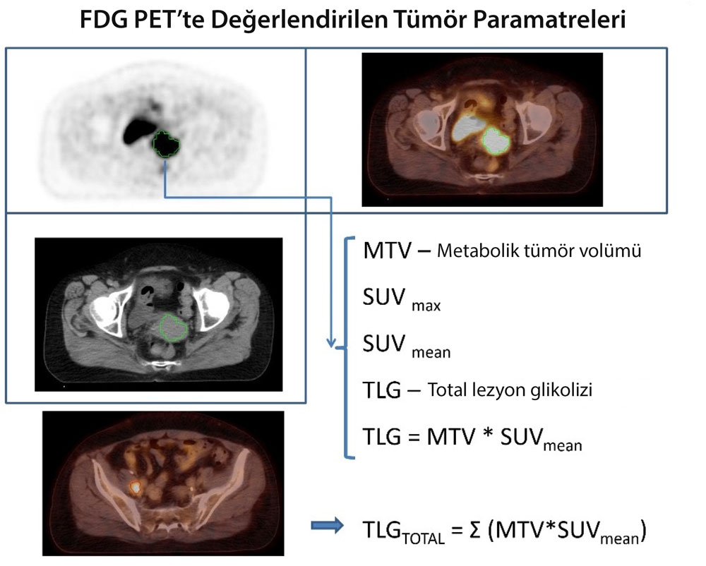
SUV (standardized uptake value) numerically measures the intensity of tissue FDG uptake. Simply put, it is the ratio of radioactivity accumulating in an area to the total administered activity per kilogram of body weight.
| Parameter | What Does It Measure? | Clinical Role |
|---|---|---|
| SUVmax | The highest single-point value in the lesion, representing the most active tumor area | The most commonly used parameter and the one emphasized in reports. Easy to measure, with good reproducibility. |
| SUVmean | All values within the lesion, expressed as their average | Reflects overall tumor activity; more variable because it depends on how boundaries are drawn. |
| MTV (metabolic tumor volume) | Tissue above a specified uptake threshold, measured by its volume | Indicates tumor burden; used in research and some lymphoma protocols. |
| TLG (total lesion glycolysis) | SUVmean × MTV: a combination of volume and intensity | Measures total metabolic burden. Prognostic value has been demonstrated, but it is less widely used than SUVmax in routine practice. |
Above What SUV Is Cancer Suspected?
A commonly used threshold is 2.5. However, this number has important limitations and is not decisive on its own.
- It Varies Between Scanners. Different PET scanners, reconstruction algorithms and centers may produce different SUVs for the same lesion. Therefore, follow-up scans should be performed at the same center when possible.
- It Depends on Timing. The longer the interval after FDG injection, generally 60 minutes, the more tumor SUV continues to rise.
- Blood Glucose Affects It. High blood glucose competes with FDG and reduces tumor uptake, potentially causing a false negative.
- It Depends on Lesion Size. In lesions smaller than approximately 8-10 mm , the partial-volume effect causes SUV to be underestimated. A small nodule's low SUV does not prove it is benign.
- Inflammation Can Easily Exceed 2.5. Active infection or sarcoidosis can produce SUVmax values of 8-10.
Which Cancers Show FDG Uptake, and Which May Not?
This distinction is important because it determines when PET is useful.
| Cancers with Intense FDG Uptake | Cancers That May Show Low FDG Uptake |
|---|---|
|
|
Why Is FDG-PET Less Effective in the Brain and Liver?
These organs share a particular problem: background noise.
What Are PET-CT and PET-MRI?
PET-CT combines positron emission tomography and computed tomography in one scanner. The principle is simple: PET identifies activity but shows anatomy poorly; CT shows anatomy well but cannot show that activity. Overlaying them identifies exactly which organ or lymph node contains a bright focus. Today, when someone says they had a PET scan, they almost always mean PET-CT.
PET-MRI combines PET with magnetic resonance imaging. It distinguishes soft tissues much better than CT and has a substantially lower ionizing radiation dose , making it advantageous for children and young people needing repeated scans. It offers advantages in the brain, liver, pelvis, and head and neck. However, scanners are less available, scans take longer, and it is inferior to CT for the lungs.
Preparing for PET: A Direct Effect on Results
PET is particularly sensitive to patient preparation. Failing to follow these rules may require a repeat scan or lead to misinterpretation.
| Rule | Reason |
|---|---|
| Fast for 4-6 hours before scanning | Eating increases glucose and insulin; insulin directs FDG into muscle and fat, away from the tumor. Only water is allowed. |
| Blood glucose is checked | High glucose competes with FDG and reduces tumor uptake. Scans are generally postponed above 150-200 mg/dL. Patients with diabetes must discuss medication and insulin timing with the center. |
| Avoid strenuous exercise for 24 hours | Active muscle takes up FDG, creating diffuse and confusing uptake. |
| Stay warm and avoid feeling cold | Cold activates brown fat, producing symmetrical neck and shoulder uptake that can mimic metastases. This is why waiting rooms are kept warm before scanning. |
| Do not talk or chew gum while waiting | The vocal cords and chewing muscles take up FDG. |
| Drink plenty of water and urinate immediately before scanning | FDG is excreted in urine; intense bladder activity can obscure pelvic lesions. |
| Report your medications | Particularly important are metformin (which causes intense bowel uptake and may temporarily be withheld on medical advice when abdominal assessment is needed), corticosteroids and bone marrow-stimulating injections . |
| Report recent procedures | Dates of surgery, biopsy, vaccination and radiotherapy directly affect interpretation. |
PET for Assessing Treatment Response
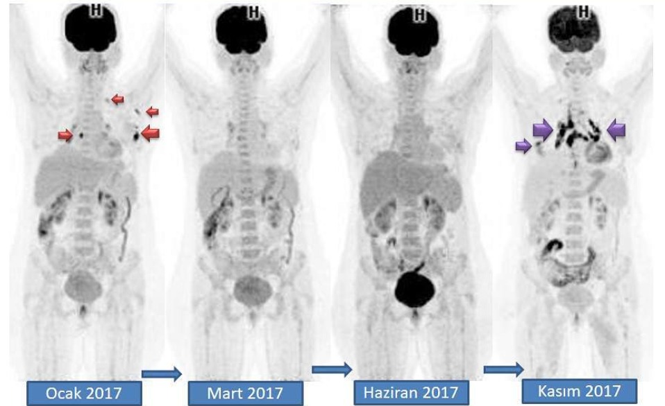
This may be its most valuable role. When a tumor responds to chemotherapy, metabolic activity falls before its size decreases —allowing PET to detect response weeks before CT can. PET also helps determine whether a residual mass is viable tumor or dead scar tissue when CT cannot.
PET Radiopharmaceuticals Other Than FDG
FDG is the most common PET tracer, but not the only one. In some cancers, imaging focuses not on the tumor's sugar consumption but on a specific target on its surface , providing much greater sensitivity.
Gallium-68 DOTATATE: Neuroendocrine Tumors

Well-differentiated neuroendocrine tumors grow slowly and have low FDG uptake , so conventional PET can be misleading. In contrast, their surfaces contain abundant somatostatin receptors , which DOTATATE binds specifically.
Gallium-68/F-18 PSMA: Prostate Cancer

PSMA (prostate-specific membrane antigen) is present in normal prostate tissue, but in prostate cancer cells it is much more abundant . By targeting this protein, PSMA-PET detects prostate cancer cells with high sensitivity.
FAPI PET
FAPI (fibroblast activation protein inhibitor) PET targets activated fibroblasts around the tumor rather than cancer cells themselves. Tumors reprogram their supporting tissue, causing these cells to produce large amounts of FAP.
FAPI has two advantages over FDG: no fasting is required and background uptake in normal tissues is very low , particularly in the brain, liver and gastrointestinal tract. It is therefore promising where FDG is less effective: liver, pancreatic, gastric and biliary cancers and peritoneal spread. For most indications it remains investigational and has not entered routine use.
| PET Tracer | Target | Principal Uses |
|---|---|---|
| F-18 FDG | Sugar (glucose) metabolism | Lung, lymphoma, colorectal, esophageal, head and neck, melanoma: the most widely used tracer |
| Ga-68 DOTATATE | Somatostatin receptors | Well-differentiated neuroendocrine tumors; eligibility for Lu-177 treatment |
| Ga-68/F-18 PSMA | Prostate-specific membrane antigen | Prostate cancer staging, localization when PSA rises, Lu-177 PSMA eligibility |
| FAPI | Activated fibroblasts surrounding the tumor | Liver, pancreatic, gastric and biliary cancers; peritoneal spread (largely investigational) |
| F-18 NaF | Bone formation activity | Bone metastases |
Related topic: An excellent treatment response wins oncology's 2018 best image award
Frequently Asked Questions
- My Report Says "FDG Uptake Observed." Do I Have Cancer?
- No. This alone does not diagnose cancer. Inflammation, infection, previous surgery, vaccination or brown fat may also cause uptake. Diagnosis requires the lesion's location and appearance, clinical history and often biopsy . Your treating physician should interpret the report.
- My SUVmax Is 6. Is That High?
- It means nothing in isolation. The same number can occur in a healing surgical wound, active infection or tumor. SUV varies with the scanner, timing, blood glucose and lesion size. What matters is interpreting the finding in its full context, not the number alone.
- Does a Clear PET Mean I Do Not Have Cancer?
- It is often reassuring but not a guarantee. Lobular breast cancer, mucinous tumors, well-differentiated neuroendocrine tumors, prostate cancer and some liver tumors may be PET-silent. In addition, lesions smaller than 8-10 mm may be missed because they are near the scanner's resolution limit.
- Can I Have My Follow-Up PET at a Different Center?
- If possible, using the same center is preferable. Different scanners and processing methods may produce different SUVs for the same lesion, making it difficult to determine whether uptake increased or decreased.
- Is PET Harmful? Can I Have It as a Check-Up?
- A PET-CT delivers approximately 10-25 mSv. This is acceptable for cancer diagnosis, staging and follow-up. However, PET is not used to screen healthy people because of unnecessary radiation and the risks of unnecessary biopsies and anxiety from false positives.
- I Have Diabetes. Can I Undergo PET?
- Yes, but preparation requires more care. Glucose must be within the target range during scanning, and medication/insulin timing must be discussed with the center. Also report if you take metformin , which causes intense bowel uptake and may hinder abdominal assessment.
- Can I Have PET Immediately After Surgery or Radiotherapy?
- Generally not. Healing-related inflammation causes intense uptake that may be mistaken for disease. After surgery, a rough waiting interval of 4-6 weeks is recommended, compared with 8-12 weeks after radiotherapy. Your physician determines the correct timing.
Sources
2. M Weineisen, M Schottelius, J Simecek, RP Baum, Akın Yıldız, et al. 68Ga-and 177Lu-labeled PSMA I&T: optimization of a PSMA-targeted theranostic concept and first proof-of-concept human studies. Journal of Nuclear Medicine, 2015
3. Czernin, J., Allen-Auerbach, M., & Schelbert, H. R. Improvements in cancer staging with PET/CT: literature-based evidence as of September 2006. Journal of Nuclear Medicine 2007
Was this article helpful?
Your feedback helps us improve our content.



Reader comments
Share your comment
Your name and comment are published only after administrator approval. Your email address is never shown to visitors.
Be the first to comment.