
PARP Inhibitors May Enhance the Power of Immunotherapy in Lung Cancer

In this article
Immunotherapy has significantly prolonged survival in many cancer types. However, only % 20 of patients respond to immunotherapies. Recently, research aimed at increasing their effectiveness has grown. In this context, in March 2019, The Journal of Clinical Investigation published a study discovering that drugs called PARP inhibitors, when used in cancer cells with defective DNA repair , may increase immune responses and expand the effectiveness of immunotherapy.
The research was conducted at the Institute of Cancer Research in London.
Researchers analyzed defects in DNA repair in tumor samples obtained from lung cancer patients. According to the research results,
- tumors with abnormalities in DNA repair genes had greater infiltration (invasion) by immune system cells. This suggests that mutations in DNA repair genes enable an immune response against tumors.
- The effects of PARP inhibitors on immune responses were also examined in cells with defects in DNA repair genes such as ERCC1 or BRCA1, including non-small cell lung cancer and triple-negative breast cancer cells.
- When cancer cells with defective DNA repair systems were treated with PARP inhibitors, they were unable to perform any DNA repair, and DNA damage accumulated until the cells died.
- The accumulation of DNA damage in cancer cells following PARP inhibitor use leads to the release of signaling molecules. These molecules stimulate immune cells to attack the tumor.
In patients with non-small cell lung cancer, % 30 – 50 have defects in the ERCC1 DNA repair system. This discovery may pave the way for effective treatments in these patients.
PARP inhibitors (for example, olaparib) block one of the systems cancer cells use to repair DNA. These inhibitors are specifically designed to target tumors with defective DNA repair systems. They are particularly effective in patients with breast and ovarian cancer carrying inherited BRCA1 mutations.
What does this study mean for us?
- PARP inhibitors were discovered to enhance the effectiveness of cancer immunotherapy. Combining PARP inhibitors and immunotherapy in cancer cells with faulty DNA repair mechanisms may increase immune responses against cancer cells.
- This approach is planned for evaluation this year in a clinical trial involving patients with lung, prostate, and bladder cancer.
*
ADVANCED INFORMATION FOR THOSE WHO WANT TO KNOW MORE: Below, the study’s graphical summary can be seen:
Modeling ERCC1 defect-dependent cGAS/STING activation following PARP inhibitor treatment
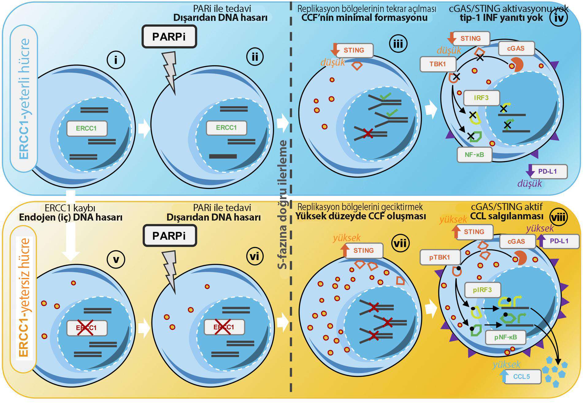
(i) ERCC1WT/WT cells have normal DNA repair systems and maintain their genomic structures.
(ii) Upon exposure to PARP inhibitors (PARPi), exogenous DNA damage is triggered, mostly initiated by PARP1 itself, located on DNA at spontaneous single-strand break sites (SSBs).
(iii) During S phase of the cell cycle, truncated PARP1 produces lesions that prevent replication forks from advancing, causing fork stalling and subsequent DSB formation. In ERCC1WT / WT cells, most trapped PARP1 lesions are removed, enabling DSB processing through HR and eventually replication restart. Residual poorly repaired lesions cause moderate CCF formation.
(iv) The low CCF levels produced cannot trigger pTBK1 / IRF3 / NF-KB signaling pathways or subsequent transcription of type-I IFN genes; PD-L1 is moderately induced.
(v) ERCC1 - / - cells are subjected to increased endogenous DNA damage following ERCC1 loss. This keeps CCF levels at a minimum.
(vi) After PARP inhibitor treatment, ERCC1 - / - cells are subjected to an additional source of exogenous DNA damage.
(vii) During S phase of the cell cycle, truncated PARP1 creates lesions that prevent replication forks from progressing, leading to fork stalling and subsequent DSB formation. In the absence of ERCC1, trapped PARP1 lesions cannot be adequately resolved, increasing DSBs and ultimately producing high CCF levels. CCFs are detected by cGAS, and because of increased STING expression in ERCC1 - / - cells, they effectively activate cGAS / STING signaling. Activated STING homodimer, through IRF3 and NF-κB phosphorylation by TBK1 which it phosphorylates, triggers their translocation into the nucleus and results in transcription of type-1 IFN genes: CCL5 and other type-I IFN cytokines are secreted. Higher PD-L1 expression is induced at the cell surface. CCFs are detected by cGAS, and because of increased STING expression in ERCC1 - / - cells, they effectively activate cGAS / STING signaling. Activated STING homodimer, through IRF3 and NF-κB phosphorylation by TBK1 which it phosphorylates, triggers their translocation into the nucleus and results in transcription of type-1 IFN genes: CCL5 and other type-1 IFN cytokines are secreted. Higher PD-L1 expression begins to appear at the cell surface.
Sources
PARP inhibition enhances tumor cell-intrinsic immunity in ERCC1-deficient non-small cell lung cancer.
J Clin Invest. 1 March 2019
Was this article helpful?
Your feedback helps us improve our content.




Reader comments
Share your comment
Your name and comment are published only after administrator approval. Your email address is never shown to visitors.
Be the first to comment.