
Thymoma and Thymic Carcinoma: Symptoms, Diagnosis and Treatment

In this article
What Are Thymoma and Thymic Carcinoma?
Key Points About Thymoma and Thymic Carcinoma:
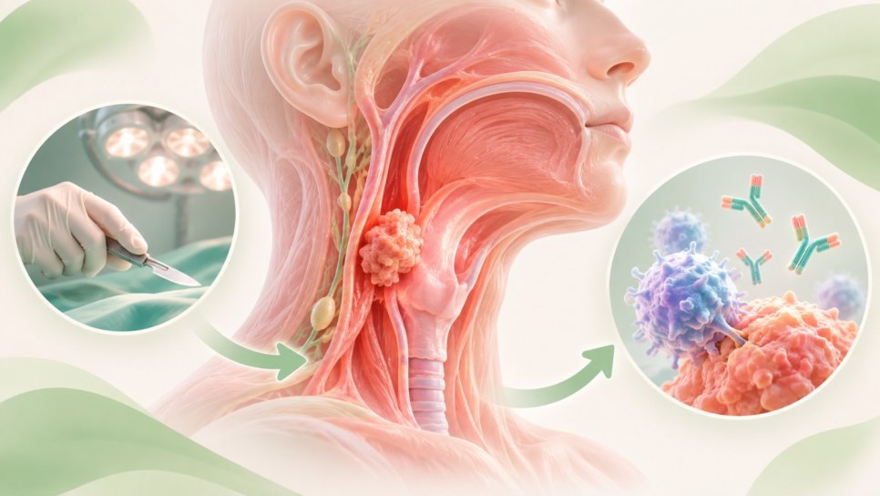
- Thymoma and thymic carcinoma are diseases in which malignant (cancer) cells are present on the outer surface of the thymus (the gland beneath the neck).
- Thymoma is associated with myasthenia gravis and other autoimmune diseases.
- The most common signs and complaints (symptoms) caused by thymoma and thymic carcinoma are cough and chest pain.
- Surgery is the main treatment for thymoma and thymic carcinoma.
- Tumor cells in thymoma resemble normal thymus cells; in medical terminology, this is called showing less atypia . In contrast, tumor cells in thymic carcinoma look very different from normal thymus cells, grow faster, and have generally spread to other parts of the body when the cancer is found. Thymic carcinomas are also called "type C thymoma." Thymic carcinoma is more difficult to treat than thymoma.
- Thymomas and thymic carcinomas account for approximately 0,2% to 1,5% of all cancers and are relatively rare tumors. According to United States cancer statistics, the overall incidence of thymoma per 100.000 people is 0.15 cases annually. Thymic carcinomas are even rarer, reportedly accounting for only 6% of all thymus gland cancers. In general, thymomas are persistent tumors that tend toward local recurrence (regional return) rather than metastasis. Thymic carcinomas, however, are typically invasive and carry a higher risk of relapse and death.
- The etiology/causes of these tumors are unknown. In approximately 50% of patients, thymomas / thymic carcinomas were detected incidentally on a plain chest X-ray.
- Most patients with thymoma or thymic carcinoma are aged 40-60.

- The thymus is a small organ in the upper chest, positioned below the upper notch of the breastbone. It is part of the lymphatic system and therefore the immune system. Its main function is producing immune cells called lymphocytes, a type of white blood cell.
- 90% of thymomas and thymic carcinomas are in the front part of the chest, called the anterior mediastinum. They are the most common cancers of this region.
Thymoma Is Associated with Myasthenia Gravis and Other Autoimmune Diseases.
People with thymoma often also have autoimmune diseases. These diseases cause the immune system to attack healthy tissues and organs.
These Autoimmune Diseases Include:
- Myasthenia gravis.
- Acquired pure red cell aplasia.
- Hypogammaglobulinemia.
- Polymyositis.
- Lupus erythematosus.
- Rheumatoid arthritis.
- Thyroiditis.
- Sjögren syndrome.
The Most Common Signs and Complaints Caused by Thymoma and Thymic Carcinoma Are Cough and Chest Pain.
Thymoma and thymic carcinoma may not cause signs or symptoms early on. The cancer may be found during a routine chest X-ray. These signs and symptoms may be caused by thymoma, thymic carcinoma or other health problems.
Consult Your Doctor If You Have Any of the Following:
The Following Tests and Procedures May Be Used to Diagnose Thymoma and Thymic Carcinoma:
Physical Examination and Patient History: An examination to check the body's general health, for example for a palpable mass. The patient's history of health habits, previous illnesses and treatments is also recorded.
Chest X-ray: An X-ray image provides a rough overview of the organs in the chest.
Computed Tomography (CT) Scan: A procedure that can produce detailed images of regions inside the body, for example images of the chest taken from different angles. The images are produced by a computer connected to an X-ray machine. A dye-like contrast agent may be injected into a vein or swallowed to help organs or tissues appear more clearly. This procedure is called computed tomography (CT).
Magnetic Resonance Imaging (MRI): A procedure using a magnet, radio waves and a computer to produce detailed images of areas inside the body, such as the chest. MRIprovides particularly good imaging of soft tissues.
Positron Emission Tomography (PET scan): A procedure for finding malignant (cancerous) tumor cells. A small amount of radioactive glucose (sugar) is injected into a vein. PET scanners rotate around the body and produce images showing where glucose is used in the body. Malignant tumor cells appear more prominently because they are more active and take up more glucose than normal cells.
To diagnose the disease, a tissue sample is taken from the tumor (biopsy) and examined under a microscope in the laboratory by a pathologist. A thymus biopsy may be performed before surgery using fine-needle aspiration or a thick needle (core biopsy). A biopsy may also be taken during mediastinoscopy or mediastinotomy surgery. If the pathologist diagnoses thymoma or thymic carcinoma, they determine the type of cancer cell in the tumor.
The Following Factors Determine Prognosis (Disease Course and Outcomes) for Thymoma and Thymic Carcinoma
- Cancer stage
- Type of cancer cell
- Whether the tumor can be completely removed surgically
- The patient's general health
- Whether the cancer is newly diagnosed or has recurred (returned).
Disease Stages in Thymoma or Thymic Carcinoma
Stage I
In Stage I, cancer is found only inside the thymus. All cancer cells are within the capsule (sac) surrounding the thymus.
Stage II
In Stage II, cancer has spread through the capsule into fatty tissue around the thymus or inside the chest cavity.
Stage III
In Stage III, cancer has spread to nearby organs in the chest, including the lungs, the sac around the heart or major blood vessels carrying blood to the heart.
Stage IV
Stage 4 is divided into stage 4A and stage 4B, depending on where the cancer has spread.
4A is the stage in which cancer has spread extensively around the lungs and heart.
4Bis the stage in which cancer has spread to the blood or lymphatic system.
At diagnosis, thymic carcinomas have generally metastasized to other parts of the body. The staging system used for thymomas is sometimes used for thymic carcinomas.
Recurrent (Relapsed) Thymoma and Thymic Carcinoma
Recurrent thymoma and thymic carcinoma are cancers that recur (can return) after treatment. Cancer may return in the thymus or elsewhere in the body. Thymic carcinomas frequently recur. Thymomas may never recur or may recur after a long interval. The risk of another cancer type also increases after having a thymoma. For these reasons, lifelong follow-up is required.
How Are Thymoma and Thymic Carcinoma Treated?
Four Types of Standard Treatment Are Used for Patients with Thymoma and Thymic Carcinoma:
- Surgery
- Radiotherapy
- Chemotherapy
- Hormone therapy
Clinical trialstest new types of treatment.
Surgery for Thymoma and Thymic Carcinoma
Surgery to remove the tumor is the most commonly used treatment for thymoma.
Even if the doctor removes all visible cancer during surgery, some patients may receive radiation therapy afterward to kill any remaining cancer cells. Treatment given after surgery to reduce the risk of cancer returning is called adjuvant therapy .
Radiation Therapy
Radiotherapyis a cancer treatment that uses high-energy X-rays or other types of radiation to kill cancer cells or prevent their growth. There are two types of radiation therapy:
External radiation therapy uses a machine outside the body to direct radiation at the cancer.
Internal radiation therapy (brachytherapy) uses radioactive needles, seeds, wires or catheters placed inside or near the cancer.
External radiotherapy is one of the commonly preferred methods for treating thymoma and thymic carcinoma.
Chemotherapy
Chemotherapyis a cancer treatment that stops cancer cell growth by either killing the cells or stopping cell division (mitosis). When chemotherapy is taken orally or injected into a vein or muscle, the drugs enter the bloodstream and can reach cancer cells throughout the body (systemic chemotherapy).
Chemotherapy may be used to shrink the tumor before surgery or radiation therapy. This is called neoadjuvant chemotherapy.
Hormone Therapy
Hormone therapy is a cancer treatment that eliminates hormones or blocks their effects and stops cancer cell growth. Hormones are substances produced by glands in the body and circulating in the bloodstream. Some hormones can cause certain cancers to grow. If tests are positive for receptors to which hormones can bind, reducing hormone production or blocking their action is used for hormone suppression treatment.
Hormone therapy with drugs called corticosteroids may be used to treat thymoma or thymic carcinoma.
Treatment of Thymoma and Thymic Carcinoma by Stage
Treatment of Stage I and Stage II Thymoma
- Treatment of Stage I thymoma is surgery.
- Treatment of Stage II thymoma is surgery followed by adjuvant radiation therapy.
For stage 1 thymoma, complete surgical excision (removal by surgery) can provide excellent long-term survival. Adjuvant radiation therapy does not appear to provide additional benefit after complete removal of thymoma tumors confined within the capsule. In patients with stage II thymoma and pathologically demonstrated capsular invasion (spread), adjuvant radiation therapy after complete surgical excision has been accepted as standard treatment.
The largest study of early-stage thymoma reported to date involved 1320 Japanese patients. In this study, patients with stage I thymoma were treated with surgery alone, while patients with stage II thymoma received surgery and additional radiation therapy. Prophylactic (preventive) mediastinal radiation therapy did not prevent local recurrence (regional return) in patients whose stage II thymoma was completely removed surgically. Therefore, the role and risks of adjuvant radiation therapy in patients with completely removed stage II thymoma require further study.
Treatment of Stage III and Stage IV Thymoma
Stage III and Stage IV Thymoma Tumors Can Be Completely Removed Surgically, and Postoperative Treatment Options Are:
- Surgery with or without radiotherapy
- Neoadjuvant chemotherapy followed by surgery with or without radiation therapy.
Stage III thymoma can be difficult to identify before surgery, because infiltrative spread into neighboring organs may only be identified during mediastinal exploration (surgical examination of the chest cavity). Patients generally undergo aggressive surgical resection with wide margins, and adjuvant radiation therapy is considered. Invasion (spread) into other regional organs may be visible on pretreatment computed tomography imaging. Tumor-shrinking (neoadjuvant) treatment combined with chemotherapy, followed by surgery and / or radiation therapy, may be recommended (three-modality treatment).
In the Japanese study of 1320 patients, those with stage III thymoma received surgery and additional radiation therapy. Patients with stage IV thymoma were treated with radiation therapy or chemotherapy. For patients with stage III or stage IV thymoma, 5-year survival rates were 93% after complete surgical removal, 64% after subtotal resection and 36% in patients whose disease could not be treated. Prophylactic mediastinal radiation therapy did not prevent regional recurrence in patients with completely removed stage III thymoma. However, radiotherapy was seen to increase the risk of cardiac side effects and secondary cancers.
The most invasive thymomas were found to be sensitive to cisplatin -based combination chemotherapy regimens. Objective response rates of 79% to 100% have been reported for cisplatin combinations in invasive thymomas, and tumor resectability rates after combination chemotherapy range from 36% to 69%.
Chemotherapy Regimens Used in Thymoma
- Cisplatin, doxorubicin and cyclophosphamide (PAC), with or without prednisone.
- Cisplatin, doxorubicin, vincristine and cyclophosphamide (ADOC).
- Cisplatin, etoposide and epirubicin.
However, particularly when major vessels are involved, similar results have been reported with preoperative radiation therapy without chemotherapy (5-year overall survival rate 77%, and at 10 years 59%).
A United States study reported that among 26 patients receiving the PAC chemotherapy regimen, the 5-year overall survival rate was 52%, followed by radiation therapy without surgery.
The role of surgical debulking (surgically reducing the tumor burden as much as possible) in patients with stage III or stage IVA disease is controversial. Studies show that long-term survival can be achieved with chemotherapy and radiation therapy alone in many patients presenting with locally advanced or even metastatic thymoma. The value of surgery may be questioned when complete surgical removal cannot be achieved.
Treatment of Stage III and Stage IV Thymoma That Cannot Be Completely Removed Surgically Includes:
- Neoadjuvant chemotherapy followed by surgery and / or radiation therapy
- Radiotherapy alone
- Chemotherapy alone
Treatment of Thymic Carcinoma
Treatment of Thymic Carcinoma That Can Be Completely Removed Surgically Includes:
- Surgery with or without radiotherapy
- A clinical trial of anticancer drugs in new combinations or doses.
- A clinical trial of new ways of delivering radiation therapy.
Treatment of Thymic Carcinoma That Cannot Be Completely Removed Surgically Includes:
- Radiation therapy.
- Surgery to remove part of the tumor, with or without chemotherapy, and/or radiotherapy.
- Chemotherapy with radiation therapy (chemoradiotherapy).
- Chemotherapy alone.
- Participation in clinical trials
Treatment of Recurrent Thymoma and Thymic Carcinoma
Treatment of Recurrent (Relapsed) Thymoma and Thymic Carcinoma May Include:
- Surgery with radiotherapy or alone.
- Radiation therapy.
- Hormone therapy.
- Chemotherapy.
- A clinical trial.
Sources
US National Cancer Institute website cancer.gov - Updated: February 9, 2018
Was this article helpful?
Your feedback helps us improve our content.
Reader comments
Share your comment
Your name and comment are published only after administrator approval. Your email address is never shown to visitors.
Be the first to comment.