
What Is PD-L1? The Importance of Testing and Positivity in Cancer Immunotherapy

In this article
Cancer cells can evade immunity by displaying abundant PD-L1 proteins on their surfaces. PD-L1 acts as a stop sign that deactivates immune T cells.
Cancer cells and the immune system interact intensely. Immune checkpoints are the molecules playing key roles in this interaction, whose discovery opened the way to cancer immunotherapies . The best-known molecules are:
- PD-1: Programmed death-1 receptor (programmed death 1),
- PD-L1: Programmed death ligand 1 (programmed death ligand 1),
- PD-L2: Programmed death ligand 2 and
- B7.1.
Why Do Our Cells Need Immune-Suppressing Molecules Like PD-L1?
The reason is simple: to prevent the immune system recognizing our own cells as foreign. However, because tumors are altered versions of our own cells, this important mechanism can become a disadvantage in cancer.
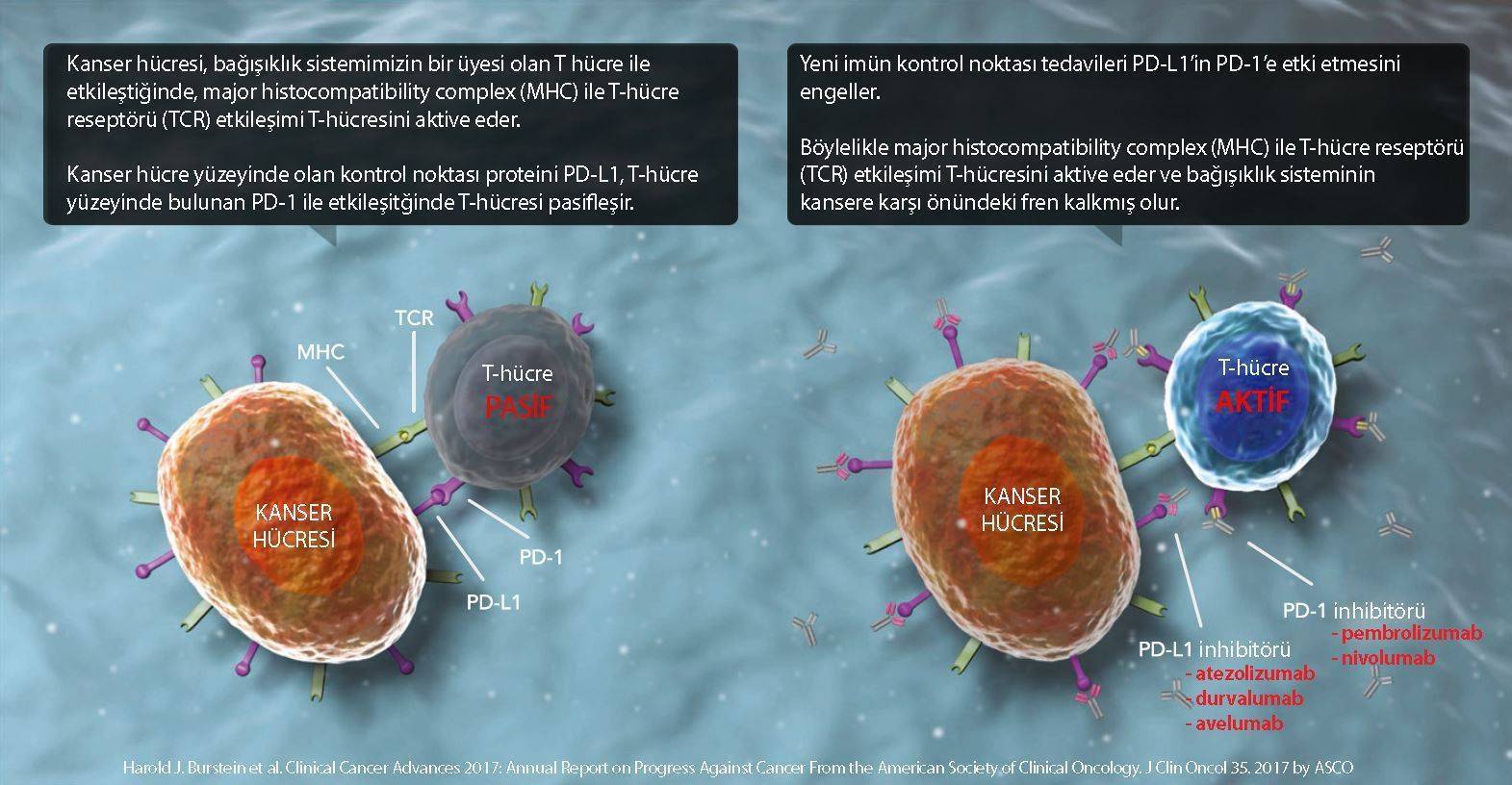
The figure belowillustrates how T cells, major immune components, do not perceive a normal epithelial cell as foreign through immune checkpoint molecules.

PD-1is a protein membrane receptor in the immunoglobulin superfamily, found on activated T cells and antigen-presenting cells.
PD-L1is a membrane protein found on cancer cells, the tumor microenvironmenton immune cells within it and on various healthy tissues, playing an important role in suppressing immunity.
PD-L2is a membrane protein on antigen-presenting immune cells.
The figure below, illustrates how T cells perceive a tumor cell as friendly following immune checkpoint interactions.

Immune checkpoint inhibitors, new-generation cancer immunotherapies, aim to activate immunity against cancer by blocking interactions such as PD-L1 and PD-1. This is achieved using monoclonal antibody medicines that block molecules deactivating immunity. However, healthy cells may also be partly affected by the increased immune response.
The figure belowdepicts monoclonal antibody immunotherapies developed against both PD-1 and PD-L1.

What Does PD-L1-Positive Mean?
Some tumors display high amounts of PD-L1 on their cell surfaces and are called PD-L1-positive tumors . PD-L1 is a valuable anti-PD-1 and anti-PD-L1 treatment-efficacy-predicting biomarkerfor these immunotherapy classes.
- FDA approved anti-PD-1 immunotherapies: pembrolizumab (marketed as Keytruda), nivolumab (Opdivo), cemiplimab (Libtayo)
- anti-PD-L1 immunotherapies: atezolizumab (Tecentriq), durvalumab (Imfinzi) and avelumab (Bevancio)
Different PD-L1 Measurement Methods
PD-L1 density differs among tumors. PD-L1 positivity is currently assessed using 3 scores.
- TPS (tumor proportion score)
- CPS (combined proportion score)
- TC (tumor cell), IC (immune cell) score
Below is how these 3 scores are calculated through immunohistochemistry (IHC) staining of pathology blocks:
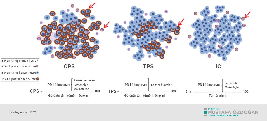
Explanation of the Figure Above: Schematic representation of current scoring criteria for PD-L1 assessment. CPS (combined positive score): the ratio of PD-L1-positive tumor and immune cells to total tumor cells. TPS (tumor proportion score): the ratio of PD-L1-positive tumor cells to all tumor cells. IC (immune cell): the ratio of PD-L1-positive immune cells to tumor area.
PD-L1 Test Kits
Different kits have been developed for different PD-L1 measurement types, and FDA (United States Food and Drug Administration), when making immunotherapy use conditional on PD-L1 positivity — not every approval requires PD-L1 positivity — specifies the required kit at approval. The table lists which PD-L1 kit should be used for each cancer and immunotherapy class.
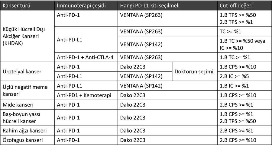
Let us explain tumor proportion score (TPS) with examples. TPS indicates the percentage of cancer cells in a pathology sample that are PD-L1-positive. According to TPS, PD-L1 positivity is classified into 3 categories:
- TPS <%1: PD-L1-negative
- TPS %1 - 49: weakly PD-L1-positive
- TPS ≥%50: strongly PD-L1-positive
For example, a study in non-small cell lung cancer (NSCLC) patients found:
- %10.6 had TPS ≥%50;
- %24.7 had TPS %1 – 49 and
- %64.7 had TPS <%1.
Tumors displaying higher PD-L1 levels are known to respond better to immune checkpoint inhibitors.

PD-L1 also has controversial aspects as a biomarker. Some PD-L1-negative patients respond well to immunotherapy, suggesting mechanisms beyond known checkpoints affect cancer-immune interactions. Another debated issue is that PD-L1 changes over time.
How Is PD-L1 Testing Done?
Testing a tumor biopsy sample for PD-L1 is recommended. Attempts to perform PD-L1 testing through liquid biopsy (fluid or blood samples) also exist, but liquid PD-L1 testing is not yet FDA-approved.
- A doctor collects a cancer biopsy and sends it to pathology. The laboratory embeds it in paraffin and cuts thin sections.
- Paraffin sections are examined using PD-L1-specific immunohistochemistry. The more PD-L1-positive the section, the darker brown the image.
- PD-L1 results are generally available within 10 working days.
- A sample must contain at least 100 tumor cells for adequate assessment.
Conclusion
PD-L1 suppresses immune T-cell activation and promotes tumor progression. In lung, stomach, liver, kidney, esophageal, pancreatic, ovarian and bladder cancers, PD-L1 overexpression or positivity is associated with poor outcomes, but in breast cancer with better outcomes. PD-L1's prognostic value in colorectal cancer and melanoma is disputed. Other biomarkers therefore take priority in colorectal cancer, such asMSI, TML and similar markers.
Studies show tumor PD-L1 levels may guide immunotherapy selection. Depending on cancer type and organ, patients with PD-L1-positive tumors show 2-3 times greater objective response to checkpoint inhibitors than negative patients.
Currently, the recommended FDA-approved PD-L1 measurement method is immunohistochemical tumor-tissue PD-L1 protein measurement. In patients lacking tissue biopsy or unable to undergo it, PD-L1 may be examined through blood-based liquid biopsy. Publications show plasma sPD-L1 rises in certain cancers and is associated with poor prognosis and metastasis . In patients with PD-L1-positive tumor tissue, plasma sPD-L1 may be a strong biomarker during immunotherapy monitoring. It should not be used alone for treatment decisions, but assessed with tumor PD-L1 levels and all other prognostic parameters.
*
RELATED TOPICS
- Oncology Terms Whose Meanings Are Often Asked
- What Are Immunotherapy Side Effects and How Are They Treated?
- What Is Microsatellite Instability? What Is DNA Mismatch Repair?
Sources
Prognostic significance of PD-L1 expression and tumor infiltrating lymphocyte in surgically resectable non-small cell lung cancer.
Oncotarget. 2017 Aug.
Was this article helpful?
Your feedback helps us improve our content.




Reader comments
Share your comment
Your name and comment are published only after administrator approval. Your email address is never shown to visitors.
Be the first to comment.